
Search Count: 53,697
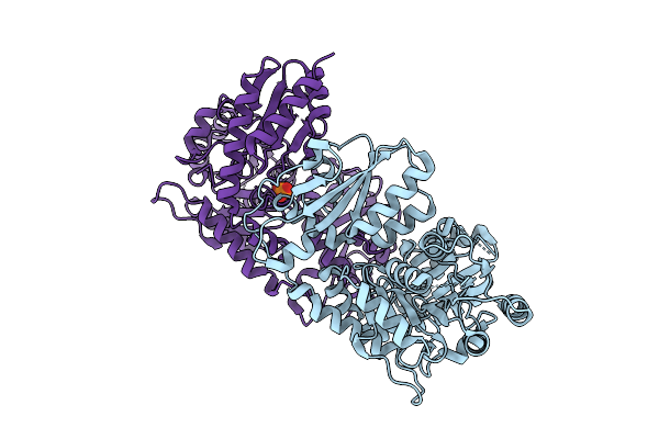 |
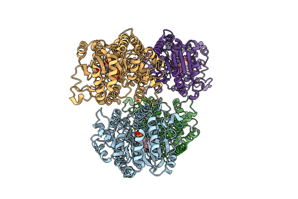
Organism: Penicillium citrinum
Method: X-RAY DIFFRACTION Release Date: 2025-11-26 Classification: LYASE Ligands: PO4, CL |
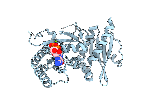 |
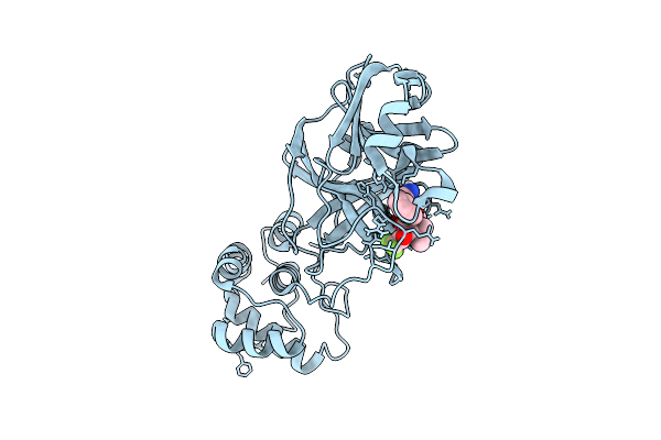
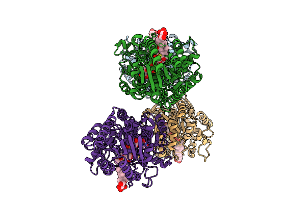
Crystal Structure Of Monomeric Rag-Like Small Gtpase From Asgard Lokiarchaeota (Lokiragm) In Complex With Gdp
Organism: Candidatus prometheoarchaeum syntrophicum
Method: X-RAY DIFFRACTION Release Date: 2025-11-26 Classification: HYDROLASE Ligands: GDP, MG |
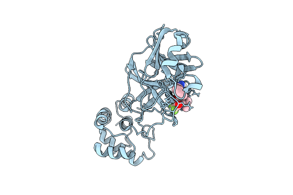 |
X-Ray Structure Of Sars-Cov-2 Main Protease V186G Covalently Bound To Compound Grl-051-22 At 1.3 A
Organism: Severe acute respiratory syndrome coronavirus 2
Method: X-RAY DIFFRACTION Release Date: 2025-11-26 Classification: VIRAL PROTEIN, HYDROLASE Ligands: A1BFE |
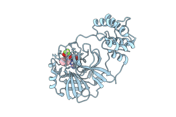 |
X-Ray Structure Of Sars-Cov-2 Main Protease Covalently Bound To Compound Grl-051-22 At 1.75 A.
Organism: Severe acute respiratory syndrome coronavirus 2
Method: X-RAY DIFFRACTION Release Date: 2025-11-26 Classification: VIRAL PROTEIN, HYDROLASE Ligands: A1BFE |
 |
X-Ray Structure Of Sars-Cov-2 Main Protease T190I Covalently Bound To Compound Grl-051-22 At 1.5 A
Organism: Severe acute respiratory syndrome coronavirus
Method: X-RAY DIFFRACTION Release Date: 2025-11-26 Classification: VIRAL PROTEIN, HYDROLASE Ligands: A1BFE, NA |
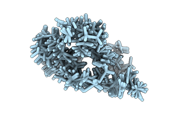 |
Staphylococcus Aureus Moaa C-Terminal Tail Peptide In Complex With G340A-Moaa
Organism: Staphylococcus aureus
Method: SOLUTION NMR Release Date: 2025-11-26 Classification: BIOSYNTHETIC PROTEIN |
 |
Organism: Orthopoxvirus vaccinia
Method: ELECTRON MICROSCOPY Release Date: 2025-11-26 Classification: TRANSCRIPTION Ligands: MG, ZN |
 |
Organism: Homo sapiens, Sars bat coronavirus
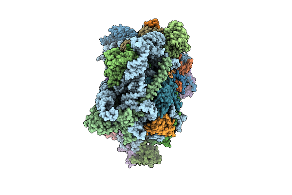
Method: ELECTRON MICROSCOPY Release Date: 2025-11-26 Classification: RIBOSOME |
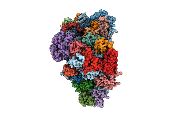 |
Organism: Homo sapiens, Sars bat coronavirus
Method: ELECTRON MICROSCOPY Release Date: 2025-11-26 Classification: RIBOSOME |
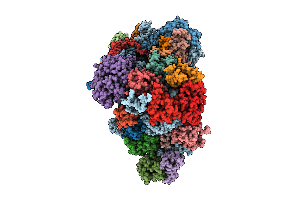 |
Organism: Homo sapiens, Sars bat coronavirus
Method: ELECTRON MICROSCOPY Release Date: 2025-11-26 Classification: RIBOSOME |
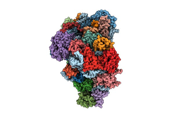 |
Organism: Homo sapiens, Sars bat coronavirus
Method: ELECTRON MICROSCOPY Release Date: 2025-11-26 Classification: RIBOSOME |
 |
Organism: Homo sapiens, Sars bat coronavirus
Method: ELECTRON MICROSCOPY Release Date: 2025-11-26 Classification: RIBOSOME |
 |
Organism: Homo sapiens, Middle east respiratory syndrome-related coronavirus
Method: ELECTRON MICROSCOPY Release Date: 2025-11-26 Classification: RIBOSOME |
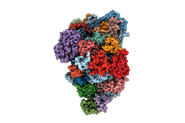 |
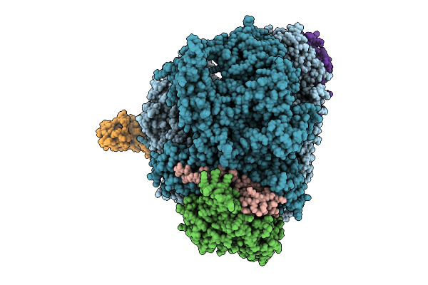
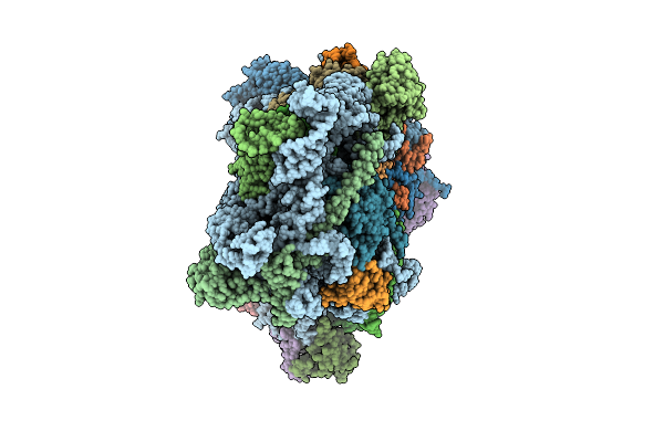
Bat Mersr-Cov Nl140422 Nsp1 Bound To The Human 40S Ribosomal Subunit-State1
Organism: Homo sapiens, Middle east respiratory syndrome-related coronavirus
Method: ELECTRON MICROSCOPY Release Date: 2025-11-26 Classification: RIBOSOME |
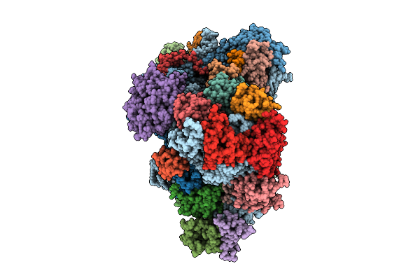 |
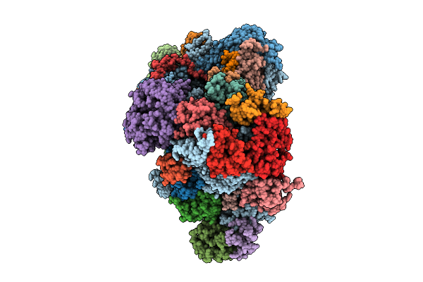
Bat Mersr-Cov Nl140422 Nsp1 Bound To The Human 40S Ribosomal Subunit-State2
Organism: Homo sapiens, Middle east respiratory syndrome-related coronavirus
Method: ELECTRON MICROSCOPY Release Date: 2025-11-26 Classification: RIBOSOME |
 |
Organism: Trifolium subterraneum
Method: X-RAY DIFFRACTION Release Date: 2025-11-26 Classification: TRANSFERASE Ligands: UDP |
 |
Organism: Trifolium subterraneum
Method: X-RAY DIFFRACTION Release Date: 2025-11-26 Classification: TRANSFERASE Ligands: A1L68, BMA, UDP |
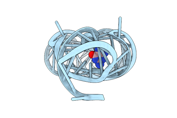 |
Organism: Paenibacillus sp. fsl e2-0178
Method: X-RAY DIFFRACTION Release Date: 2025-11-26 Classification: RNA Ligands: GUN, MG |
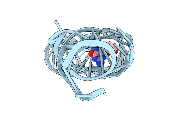 |
Crystal Structure Of Guanine-Ii Riboswitch In Complex With 2'-Deoxyguanosine
Organism: Paenibacillus sp. fsl e2-0178
Method: X-RAY DIFFRACTION Release Date: 2025-11-26 Classification: RNA Ligands: MG, GNG |
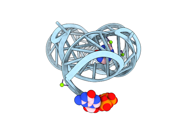 |
Organism: Paenibacillus sp. fsl e2-0178
Method: X-RAY DIFFRACTION Release Date: 2025-11-26 Classification: RNA Ligands: HPA, GTP, MG |