
Search Count: 23
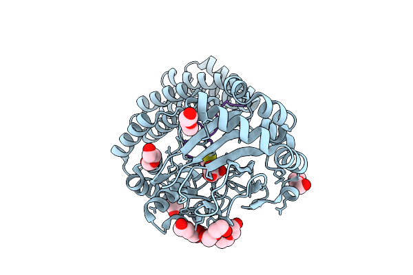 |
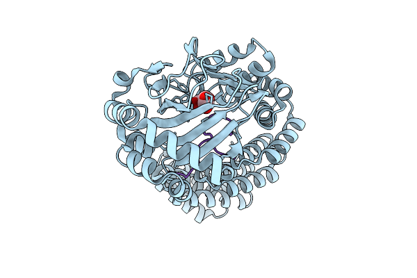
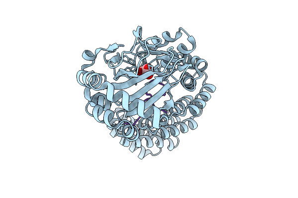
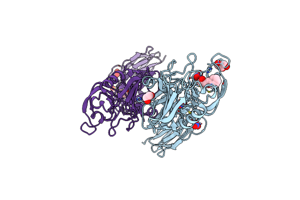
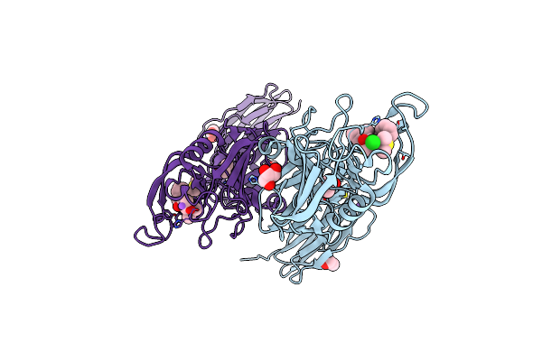
Aspartyl/Asparaginyl Beta-Hydroxylase (Asph) In Complex With Fe, 2-Oxoglutarate, Thiocyanate And Factor X Derived Peptide Fragment
Organism: Homo sapiens
Method: X-RAY DIFFRACTION Release Date: 2025-12-24 Classification: OXIDOREDUCTASE Ligands: AKG, FE, PEG, SCN |
 |
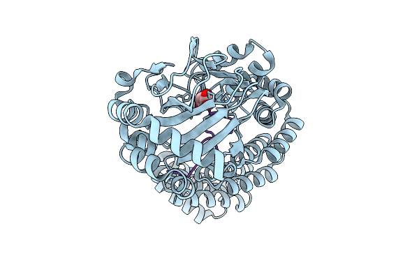
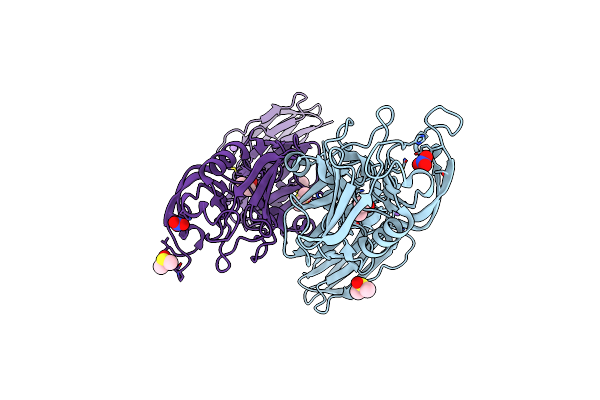
Aspartyl/Asparaginyl Beta-Hydroxylase (Asph) In Complex With Fe, 2Og, Sin And Hydroxylated Product Of Factor X Peptide Fragment (39Mer-4Ser)
Organism: Homo sapiens
Method: X-RAY DIFFRACTION Release Date: 2025-12-24 Classification: OXIDOREDUCTASE Ligands: SIN, AKG, FE |
 |
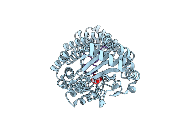
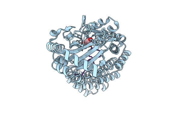
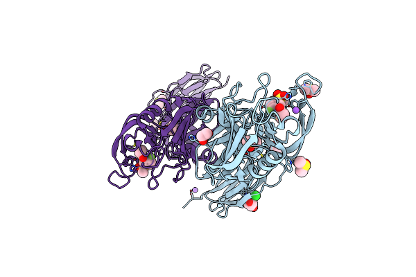
Aspartyl/Asparaginyl Beta-Hydroxylase (Asph) In Complex With Fe, 2-Oxoglutarate And Factor X Derived Peptide Fragment
Organism: Homo sapiens
Method: X-RAY DIFFRACTION Release Date: 2025-12-24 Classification: OXIDOREDUCTASE Ligands: AKG, PEG, CL, FE |
 |
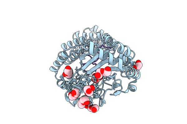
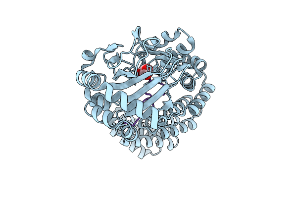
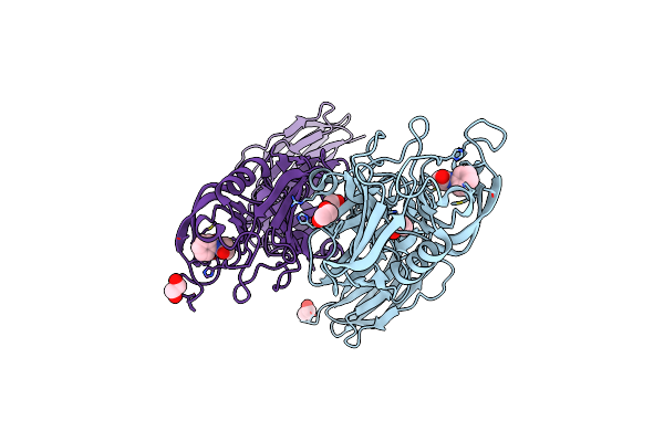
Aspartyl/Asparaginyl Beta-Hydroxylase (Asph) In Complex With Fe, 2-Oxoglutarate, Succinate And The Hudroxylated Product Of Factor X Derived Peptide Fragment After O2 Exposure
Organism: Homo sapiens
Method: X-RAY DIFFRACTION Release Date: 2025-12-24 Classification: OXIDOREDUCTASE Ligands: AKG, SIN, PEG, FE, GOL, CL |
 |
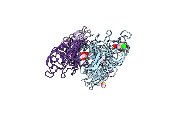
Aspartyl/Asparaginyl Beta-Hydroxylase (Asph) In Complex With Fe, 2-Oxoglutarate And A Factor X Derived Peptide Fragment
Organism: Homo sapiens
Method: X-RAY DIFFRACTION Release Date: 2025-12-24 Classification: OXIDOREDUCTASE Ligands: AKG, PEG, GOL, FE |
 |
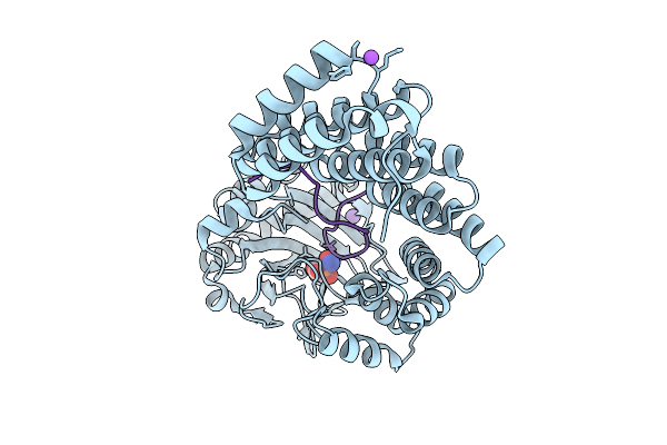
Room Temperature Structure Of Aspartyl/Asparaginyl Beta-Hydroxylase (Asph) In Complex With Fe, 2-Oxoglutarate And Factor X Derived Peptide Fragment
Organism: Homo sapiens
Method: X-RAY DIFFRACTION Release Date: 2025-12-24 Classification: OXIDOREDUCTASE Ligands: AKG, FE |
 |
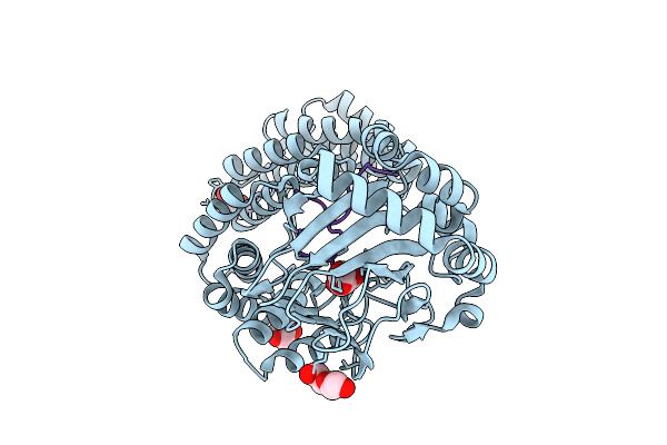
Room Temperature Structure Of Aspartyl/Asparaginyl Beta-Hydroxylase (Asph) In Complex With Fe, 2-Oxoglutarate And Hydroxylated Factor X Derived Peptide Fragment, 2 H O2 Exposure
Organism: Homo sapiens
Method: X-RAY DIFFRACTION Release Date: 2025-12-24 Classification: OXIDOREDUCTASE Ligands: SIN, FE |
 |
Aspartyl/Asparaginyl Beta-Hydroxylase (Asph) In Complex With Fe, 2-Oxoglutarate, Succinate And The Hydroxylated Product Of Factor X Derived Peptide Fragment, 12 H O2 Exposure
Organism: Homo sapiens
Method: X-RAY DIFFRACTION Release Date: 2025-12-24 Classification: OXIDOREDUCTASE Ligands: SIN, FE |
 |
Room Temperature Structure Of Aspartyl/Asparaginyl Beta-Hydroxylase (Asph) In Complex With Fe, 2-Oxoglutarate, And Hydroxylated Factor X Derived Peptide Fragment, 1.5 S O2 Exposure
Organism: Homo sapiens
Method: X-RAY DIFFRACTION Release Date: 2025-12-24 Classification: OXIDOREDUCTASE Ligands: AKG, SIN, FE |
 |
Room Temperature Structure Of Aspartyl/Asparaginyl Beta-Hydroxylase (Asph) In Complex With Fe, 2-Oxoglutarate And Factor X Derived Peptide Fragment, Excess Fe Removed
Organism: Homo sapiens
Method: X-RAY DIFFRACTION Release Date: 2025-12-24 Classification: OXIDOREDUCTASE Ligands: AKG, FE |
 |
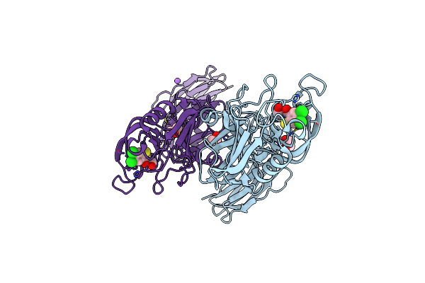
Aspartyl/Asparaginyl Beta-Hydroxylase (Asph) In Complex With Fe, 2Og, Nitric Oxide, And Factor X Peptide Fragment
Organism: Homo sapiens
Method: X-RAY DIFFRACTION Release Date: 2025-12-24 Classification: OXIDOREDUCTASE |
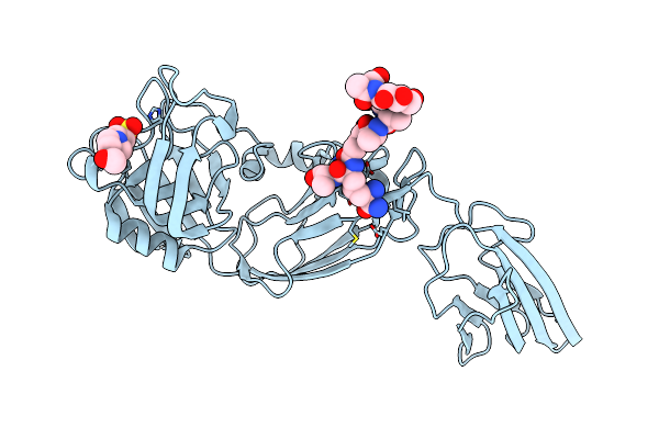 |
Crystal Structure Of The Transpeptidase Ldtmt2 C354S Mutant From Mycobacterium Tuberculosis In Complex With Natural Substrate
Organism: Mycobacterium tuberculosis, Corynebacterium jeikeium
Method: X-RAY DIFFRACTION Resolution:2.15 Å Release Date: 2024-08-07 Classification: ANTIMICROBIAL PROTEIN Ligands: EPE, CL |
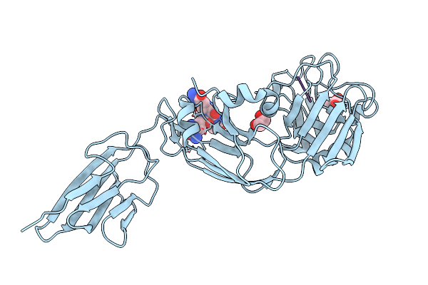 |
Crystal Structure Of The Transpeptidase Ldtmt2 From Mycobacterium Tuberculosis In Complex With Natural Substrate
Organism: Mycobacterium tuberculosis, Corynebacterium jeikeium
Method: X-RAY DIFFRACTION Resolution:1.98 Å Release Date: 2024-08-07 Classification: ANTIMICROBIAL PROTEIN Ligands: EDO, DAL, CL |
 |
Crystal Structure Of The Transpeptidase Ldtmt2 From Mycobacterium Tuberculosis In Complex With Diepoxide Ketone 1
Organism: Mycobacterium tuberculosis
Method: X-RAY DIFFRACTION Resolution:2.15 Å Release Date: 2023-11-22 Classification: ANTIMICROBIAL PROTEIN Ligands: QXU, NO3, GOL, EDO, DMS |
 |
Crystal Structure Of The Transpeptidase Ldtmt2 From Mycobacterium Tuberculosis In Complex With Cyanamide Analogue 31
Organism: Mycobacterium tuberculosis
Method: X-RAY DIFFRACTION Resolution:2.30 Å Release Date: 2023-08-02 Classification: ANTIMICROBIAL PROTEIN Ligands: NO3, DMS, NA, CL |
 |
Crystal Structure Of The Transpeptidase Ldtmt2 From Mycobacterium Tuberculosis In Complex With Maleimide Analogue 3
Organism: Mycobacterium tuberculosis
Method: X-RAY DIFFRACTION Resolution:2.55 Å Release Date: 2023-06-14 Classification: ANTIMICROBIAL PROTEIN Ligands: DMS, EDO, KSU |
 |
Crystal Structure Of The Transpeptidase Ldtmt2 From Mycobacterium Tuberculosis In Complex With Ebsulfur Analogue 15
Organism: Mycobacterium tuberculosis
Method: X-RAY DIFFRACTION Resolution:1.75 Å Release Date: 2023-06-14 Classification: ANTIMICROBIAL PROTEIN Ligands: KT6, EDO, DMS, NA |
 |
Crystal Structure Of The Transpeptidase Ldtmt2 From Mycobacterium Tuberculosis In Complex With Alpha-Chloro Ketone 2
Organism: Mycobacterium tuberculosis
Method: X-RAY DIFFRACTION Resolution:2.30 Å Release Date: 2023-06-14 Classification: ANTIMICROBIAL PROTEIN Ligands: KS9, GOL, DMS, EDO, NA, CL |
 |
Crystal Structure Of The Transpeptidase Ldtmt2 From Mycobacterium Tuberculosis In Complex With Maleimide Analogue 4
Organism: Mycobacterium tuberculosis
Method: X-RAY DIFFRACTION Resolution:2.30 Å Release Date: 2023-06-14 Classification: ANTIMICROBIAL PROTEIN Ligands: KUL, EDO, DMS, GOL, NA, CL |
 |
Crystal Structure Of The Transpeptidase Ldtmt2 From Mycobacterium Tuberculosis In Complex With Fumaryl Amide Analogue 13
Organism: Mycobacterium tuberculosis
Method: X-RAY DIFFRACTION Resolution:2.05 Å Release Date: 2023-06-14 Classification: ANTIMICROBIAL PROTEIN Ligands: KUS, GOL, DMS, NA, EDO |