
Search Count: 17
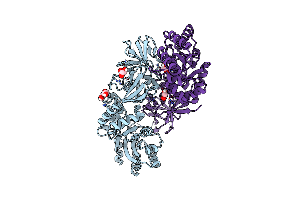 |
Structure Of Dcs-Resistant Variant D322N Of Alanine Racemase From Mycobacterium Tuberculosis
Organism: Mycobacterium tuberculosis h37rv
Method: X-RAY DIFFRACTION Resolution:1.58 Å Release Date: 2023-04-05 Classification: ISOMERASE Ligands: GOL, EDO |
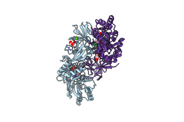 |
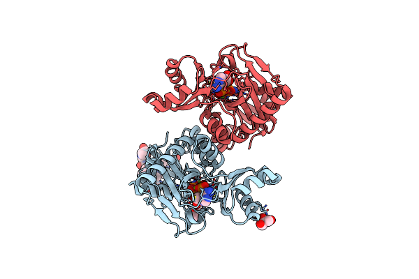
Structure Of Dcs-Resistant Variant D322N Of Alanine Racemase From M. Tuberculosis In Complex With Dcs
Organism: Mycobacterium tuberculosis h37rv
Method: X-RAY DIFFRACTION Resolution:1.78 Å Release Date: 2023-04-05 Classification: ISOMERASE Ligands: OJQ, L7N, CA, CL, GOL, EDO |
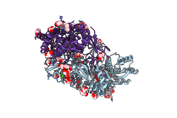 |
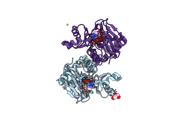
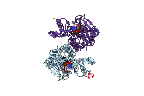
Organism: Mycobacterium tuberculosis h37rv
Method: X-RAY DIFFRACTION Resolution:1.57 Å Release Date: 2020-04-01 Classification: ISOMERASE Ligands: GOL, CA, CL, EDO, PEG, P4K, L7N, OJQ, TRS |
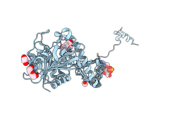 |
Organism: Mycobacterium tuberculosis (strain atcc 25177 / h37ra)
Method: X-RAY DIFFRACTION Resolution:2.26 Å Release Date: 2018-05-02 Classification: TRANSFERASE Ligands: MG, G1P, ACO, PEG, EDO |
 |
 |
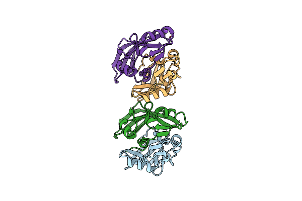
The X-Ray Crystal Structures Of D-Alanyl-D-Alanine Ligase In Complex Adp And D-Cycloserine Phosphate
Organism: Escherichia coli k-12, Synthetic construct
Method: X-RAY DIFFRACTION Resolution:1.65 Å Release Date: 2015-01-21 Classification: LIGASE Ligands: ADP, DS0, MG, GOL |
 |
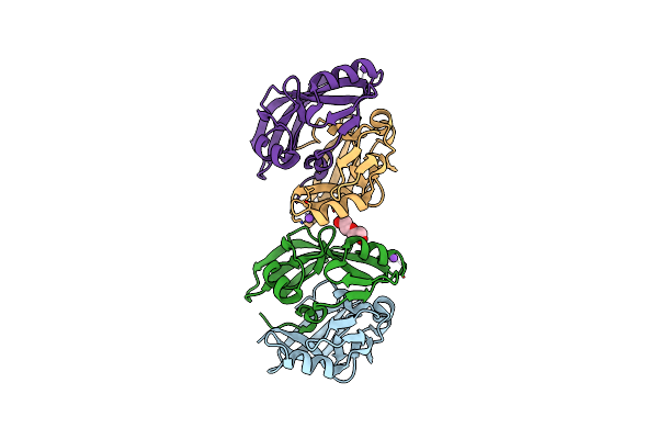
The X-Ray Crystal Structure Of D-Alanyl-D-Alanine Ligase In Complex With Atp And D-Ala-D-Ala
Organism: Escherichia coli
Method: X-RAY DIFFRACTION Resolution:1.50 Å Release Date: 2015-01-21 Classification: LIGASE Ligands: ADP, DAL, CO3, MG, GOL, IMD |
 |
The X-Ray Crystal Structure Of D-Alanyl-D-Alanine Ligase In Complex With Adp And D-Ala-D-Ala
Organism: Escherichia coli
Method: X-RAY DIFFRACTION Resolution:1.40 Å Release Date: 2015-01-21 Classification: LIGASE Ligands: ATP, DAL, MG, GOL |
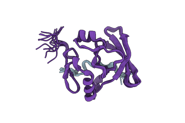 |
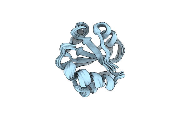
Solution Structure Of The Axh Domain Of Ataxin-1 In Complex With Ligand Peptide From Capicua
Organism: Homo sapiens
Method: SOLUTION NMR Release Date: 2013-12-11 Classification: TRANSCRIPTION REGULATOR |
 |
Organism: Homo sapiens
Method: X-RAY DIFFRACTION Resolution:2.50 Å Release Date: 2013-03-27 Classification: RNA BINDING PROTEIN |
 |
Organism: Homo sapiens
Method: X-RAY DIFFRACTION Resolution:2.45 Å Release Date: 2013-03-27 Classification: RNA BINDING PROTEIN |
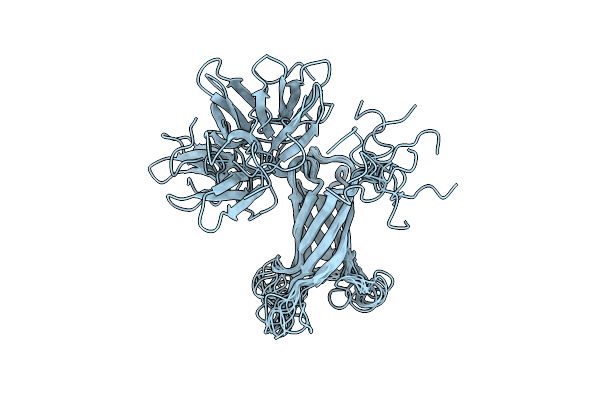 |
The Structure Of A N. Meningitides Protein Targeted For Vaccine Development
Organism: Neisseria meningitidis
Method: SOLUTION NMR Release Date: 2011-10-19 Classification: MEMBRANE PROTEIN |
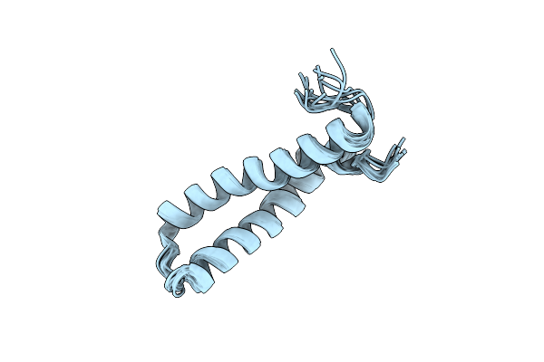 |
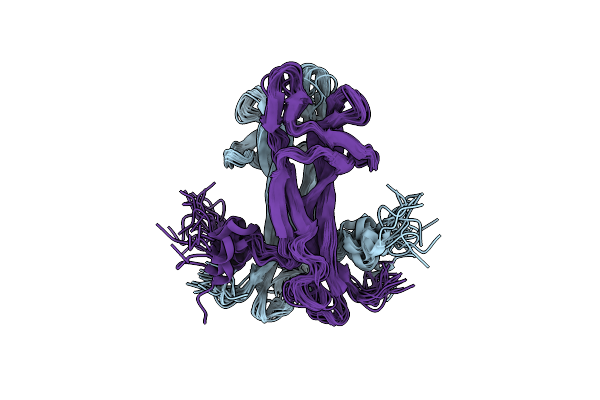
Solution Nmr Structure Of H2H3 Domain Of Ovine Prion Protein (Residues 167-234)
|
 |
Organism: Mus musculus
Method: SOLUTION NMR Release Date: 2008-04-22 Classification: GENE REGULATION, PROTEIN BINDING |
 |
Organism: Mus musculus
Method: SOLUTION NMR Release Date: 2005-04-21 Classification: DNA BINDING PROTEIN |
 |
Solution Structure Of Crotamine, A Neurotoxin From Crotalus Durissus Terrificus
Organism: Crotalus durissus terrificus
Method: SOLUTION NMR Release Date: 2003-05-09 Classification: TOXIN |
 |
Organism: Alicyclobacillus acidocaldarius
Method: SOLUTION NMR Release Date: 2000-01-26 Classification: ELECTRON TRANSPORT |