
Search Count: 20
 |
Cryo-Em Structure Of The Bcsb Hexameric Crown From The E. Coli Cellulose Secretion Macrocomplex
Organism: Escherichia coli
Method: ELECTRON MICROSCOPY Release Date: 2024-10-16 Classification: MEMBRANE PROTEIN |
 |
Cryo-Em Structure Of The C-Di-Gmp-Free Synthase:Petn Transferase Complex (Bcsa-Bct-G3) From The E. Coli Cellulose Secretion Macrocomplex
Organism: Escherichia coli
Method: ELECTRON MICROSCOPY Release Date: 2024-10-16 Classification: MEMBRANE PROTEIN |
 |
Cryo-Em Structure Of The C-Di-Gmp-Bound Synthase:Petn Transferase Complex (Bcsa-Bct-G3) From The E. Coli Cellulose Secretion Macrocomplex
Organism: Escherichia coli
Method: ELECTRON MICROSCOPY Release Date: 2024-10-16 Classification: MEMBRANE PROTEIN Ligands: C2E |
 |
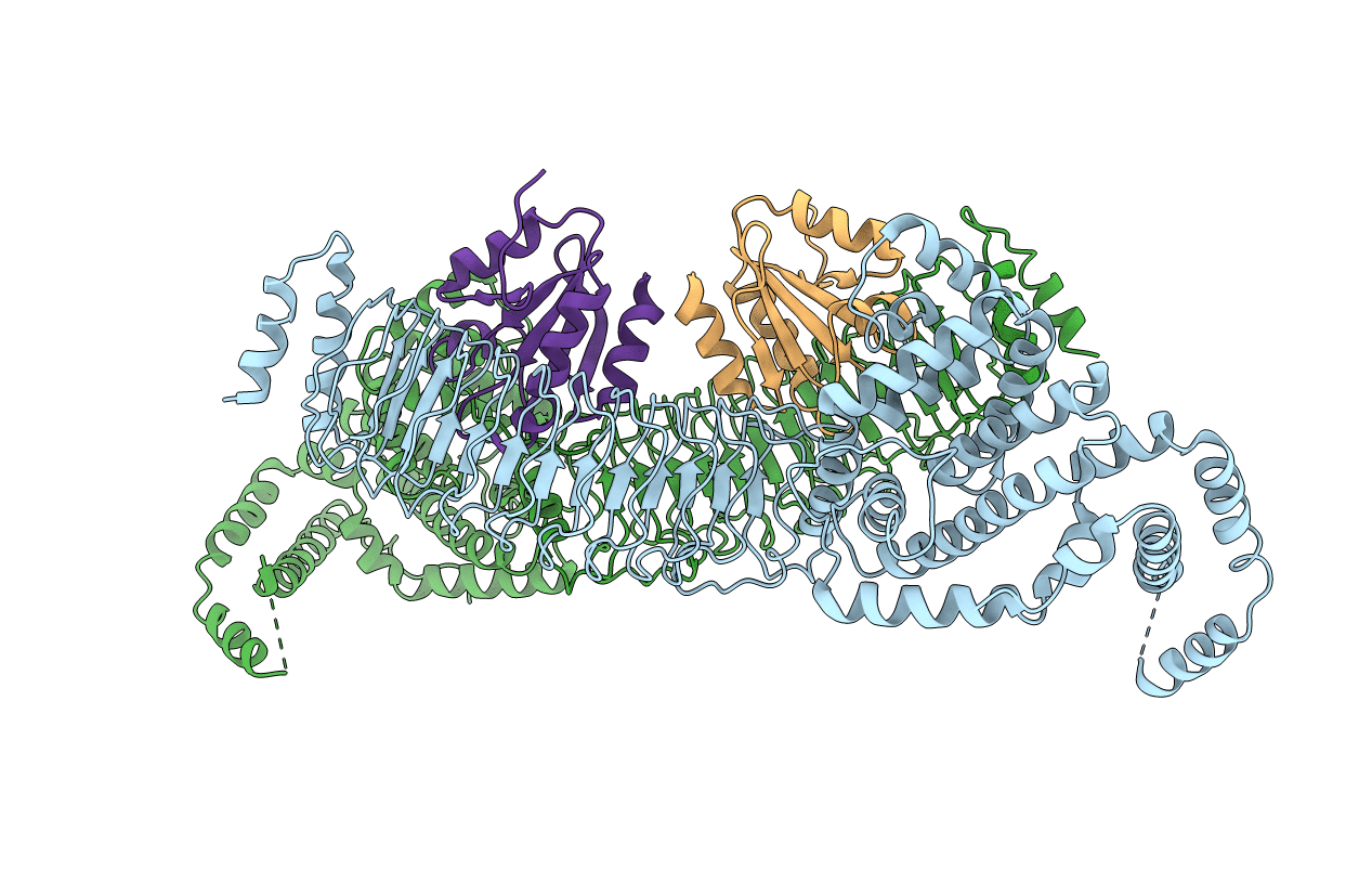
Cryo-Em Structure Of The C-Di-Gmp-Saturated 'Crown'Less Bcs Macrocomplex For Cellulose Secretion In E. Coli
Organism: Escherichia coli
Method: ELECTRON MICROSCOPY Release Date: 2024-10-16 Classification: MEMBRANE PROTEIN Ligands: C2E, ATP, MG |
 |
Cryo-Em Structure Of The Bcse2F2 Regulatory Subcomplex From The E. Coli Bcs Macrocomplex For Cellulose Secretion (Local Refinement)
Organism: Escherichia coli
Method: ELECTRON MICROSCOPY Release Date: 2024-10-16 Classification: MEMBRANE PROTEIN Ligands: C2E |
 |
Cryo-Em Structure Of The 'Crown'Less Bcs Macrocomplex For E. Coli Cellulose Secretion In Non-Saturating C-Di-Gmp (Local)
Organism: Escherichia coli
Method: ELECTRON MICROSCOPY Release Date: 2024-10-16 Classification: MEMBRANE PROTEIN Ligands: C2E, ATP, MG |
 |
Cryo-Em Structure Of The Bcsefrq Regulatory Subcomplex For E. Coli Cellulose Secretion In Non-Saturating C-Di-Gmp (Local)
Organism: Escherichia coli
Method: ELECTRON MICROSCOPY Release Date: 2024-10-16 Classification: MEMBRANE PROTEIN Ligands: C2E, ATP, MG |
 |
Organism: Pseudomonas aeruginosa (strain atcc 15692 / dsm 22644 / cip 104116 / jcm 14847 / lmg 12228 / 1c / prs 101 / pao1)
Method: X-RAY DIFFRACTION Resolution:1.74 Å Release Date: 2021-07-21 Classification: HYDROLASE Ligands: PO4 |
 |
Crystal Structure Of A Selenium-Derivatized Complex Of The Bacterial Cellulose Secretion Regulators Bcsr And Bcsq, Crystallized In The Presence Of Appcp
Organism: Escherichia coli
Method: X-RAY DIFFRACTION Resolution:1.90 Å Release Date: 2021-02-24 Classification: SIGNALING PROTEIN Ligands: MG, ATP |
 |
Crystal Structure Of A Selenium-Derivatized Complex Of The Bacterial Cellulose Secretion Regulators Bcsr And Bcsq, Crystallized In The Presence Of Adp
Organism: Escherichia coli
Method: X-RAY DIFFRACTION Resolution:2.09 Å Release Date: 2021-02-24 Classification: SIGNALING PROTEIN Ligands: MG, ATP |
 |
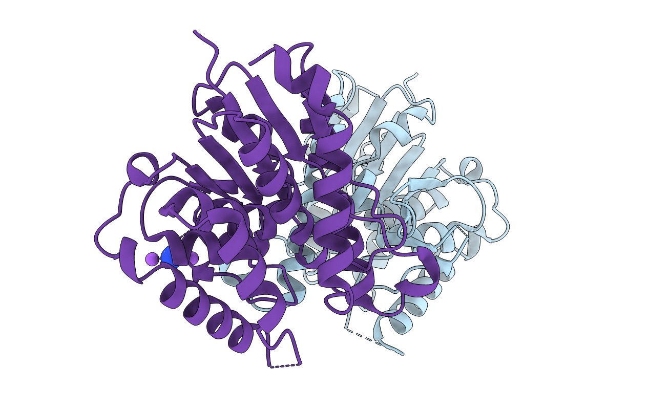
Crystal Structure Of A Native Bcsrq Complex Purified And Crystallized In The Absence Of Nucleotide
Organism: Escherichia coli
Method: X-RAY DIFFRACTION Resolution:1.59 Å Release Date: 2021-02-24 Classification: SIGNALING PROTEIN Ligands: MG, ATP |
 |
Orthorhombic Crystal Structure Of A Native Bcsrq Complex Crystallized In The Presence Of Adp
Organism: Escherichia coli
Method: X-RAY DIFFRACTION Resolution:1.59 Å Release Date: 2021-02-24 Classification: SIGNALING PROTEIN Ligands: MG, ATP |
 |
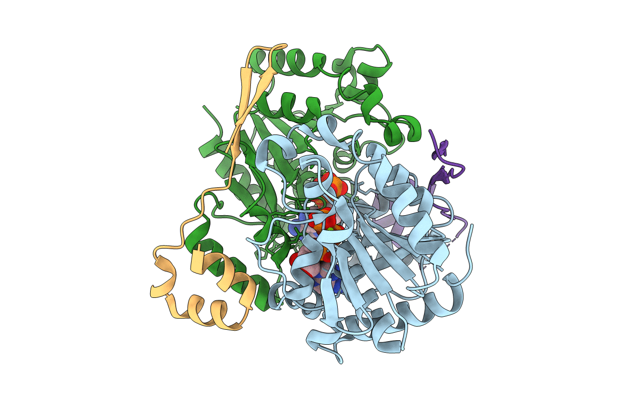
Crystal Structure Of A Native Bcse (217-523) - Bcsr-Bcsq (R156E Mutant) Complex With C-Di-Gmp And Atp Bound
Organism: Escherichia coli, Escherichia coli (strain k12)
Method: X-RAY DIFFRACTION Resolution:2.90 Å Release Date: 2021-02-24 Classification: SIGNALING PROTEIN Ligands: MG, ATP, C2E, GOL |
 |
Crystal Structure Of A Native Bcse (349-523) Rq Complex With C-Di-Gmp And Atp Bound
Organism: Escherichia coli, Escherichia coli (strain k12)
Method: X-RAY DIFFRACTION Resolution:2.49 Å Release Date: 2021-02-24 Classification: SIGNALING PROTEIN Ligands: ATP, MG, C2E |
 |
Cryo-Em Structure Of A Bcsb Pentamer In The Context Of An Assembled Bcs Macrocomplex
Organism: Escherichia coli
Method: ELECTRON MICROSCOPY Release Date: 2021-02-24 Classification: SIGNALING PROTEIN |
 |
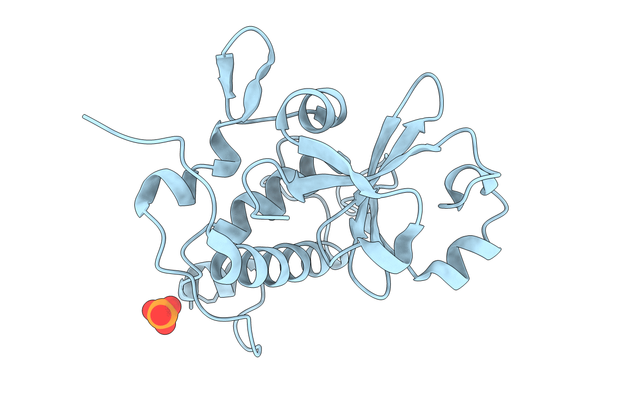
Crystal Structure Of The Bacterial Cellulose Secretion Regulator Bcse, Residues 217-523, With Bound C-Di-Gmp.
Organism: Escherichia coli k-12
Method: X-RAY DIFFRACTION Resolution:2.20 Å Release Date: 2020-07-29 Classification: SIGNALING PROTEIN Ligands: GOL, C2E |
 |
Organism: Pseudomonas aeruginosa pao1
Method: X-RAY DIFFRACTION Resolution:2.74 Å Release Date: 2016-04-06 Classification: TRANSPORT PROTEIN Ligands: AZI, NA |
 |
Organism: Pseudomonas aeruginosa
Method: X-RAY DIFFRACTION Resolution:2.14 Å Release Date: 2016-04-06 Classification: HYDROLASE |
 |
Complex Between The Salmonella T3Ss Effector Slrp And Its Human Target Thioredoxin-1
Organism: Salmonella enterica subsp. enterica serovar typhimurium, Homo sapiens
Method: X-RAY DIFFRACTION Resolution:3.30 Å Release Date: 2014-09-17 Classification: LIGASE/OXIDOREDUCTASE |
 |
Organism: Bacillus thuringiensis serovar thuringiensis
Method: X-RAY DIFFRACTION Resolution:3.20 Å Release Date: 2013-07-03 Classification: Transcription, Peptide binding protein |