
Search Count: 21
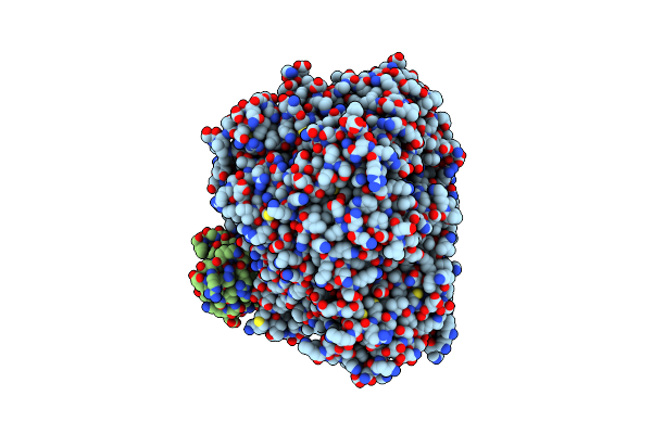 |
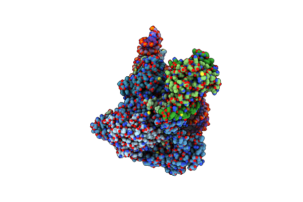
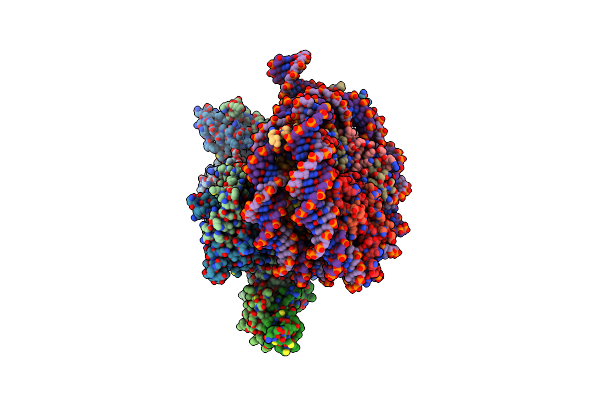
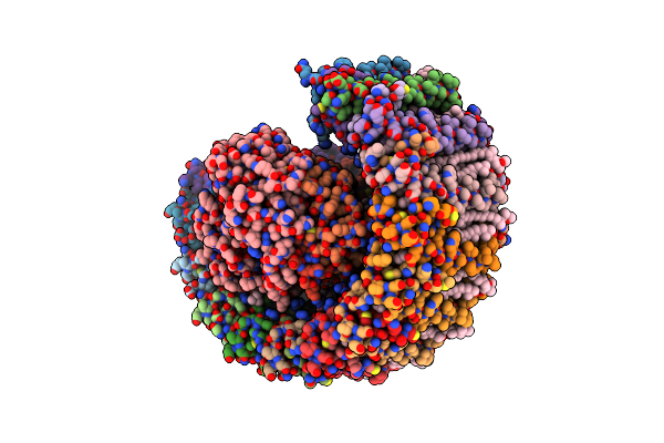
Cryo-Em Structure Of A Truncated Nipah Virus L Protein Bound By Phosphoprotein Tetramer
Organism: Henipavirus nipahense
Method: ELECTRON MICROSCOPY Release Date: 2025-05-21 Classification: VIRAL PROTEIN Ligands: ZN |
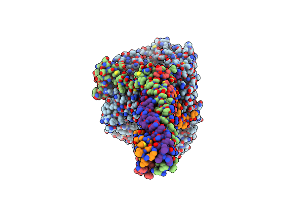 |
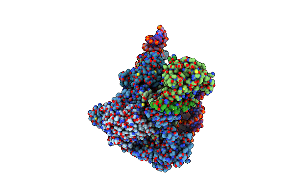
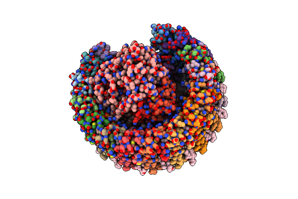
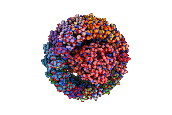
Cryo-Em Structure Of The Full-Length Nipah Virus L Protein Bound By Phosphoprotein Tetramer
Organism: Henipavirus nipahense
Method: ELECTRON MICROSCOPY Release Date: 2025-05-21 Classification: VIRAL PROTEIN Ligands: ZN |
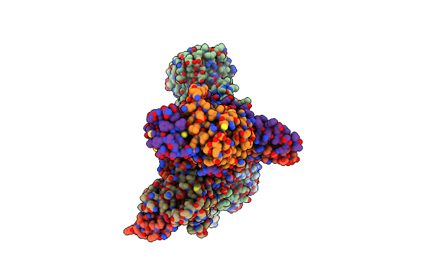 |
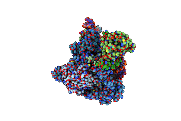
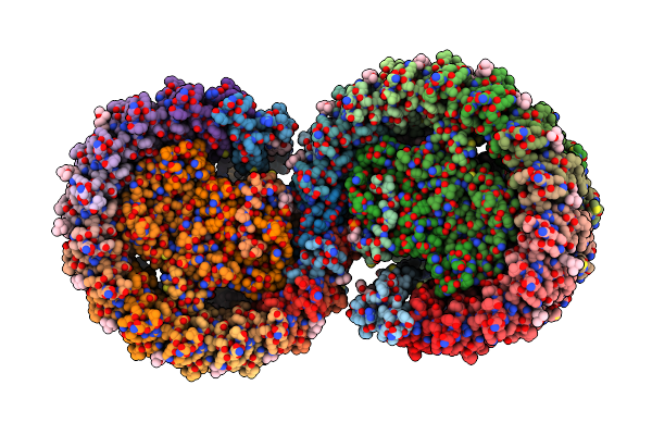
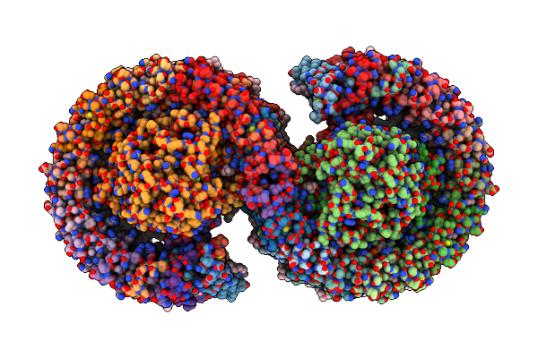
Organism: Saccharomyces cerevisiae (strain atcc 204508 / s288c)
Method: ELECTRON MICROSCOPY Release Date: 2023-09-13 Classification: TRANSCRIPTION |
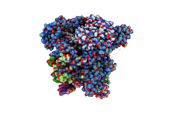 |
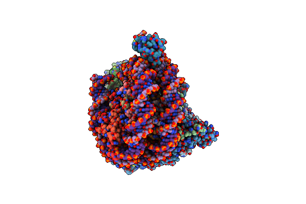
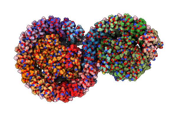
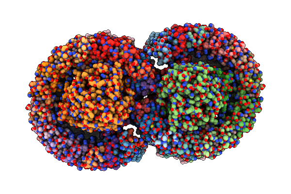
Organism: Saccharomyces cerevisiae, Xenopus laevis, Synthetic construct
Method: ELECTRON MICROSCOPY Release Date: 2023-09-13 Classification: TRANSCRIPTION |
 |
Rpd3S In Complex With Nucleosome With H3K36Mla Modification, H3K9Q Mutation And 187Bp Dna
Organism: Saccharomyces cerevisiae, Xenopus laevis, Synthetic construct
Method: ELECTRON MICROSCOPY Release Date: 2023-09-13 Classification: TRANSCRIPTION Ligands: ZN |
 |
Rpd3S In Complex With Nucleosome With H3K36Mla Modification And 187Bp Dna, Class1
Organism: Saccharomyces cerevisiae, Xenopus laevis, Synthetic construct
Method: ELECTRON MICROSCOPY Release Date: 2023-09-13 Classification: TRANSCRIPTION Ligands: ZN |
 |
Rpd3S In Complex With Nucleosome With H3K36Mla Modification And 187Bp Dna, Class2
Organism: Saccharomyces cerevisiae, Xenopus laevis, Synthetic construct
Method: ELECTRON MICROSCOPY Release Date: 2023-09-13 Classification: TRANSCRIPTION Ligands: ZN |
 |
Rpd3S In Complex With Nucleosome With H3K36Mla Modification And 187Bp Dna, Class3
Organism: Saccharomyces cerevisiae, Xenopus laevis, Synthetic construct
Method: ELECTRON MICROSCOPY Release Date: 2023-09-13 Classification: TRANSCRIPTION Ligands: ZN |
 |
Organism: Saccharomyces cerevisiae, Xenopus laevis, Synthetic construct
Method: ELECTRON MICROSCOPY Release Date: 2023-09-13 Classification: TRANSCRIPTION |
 |
Organism: Cereibacter sphaeroides 2.4.1
Method: ELECTRON MICROSCOPY Release Date: 2022-06-01 Classification: PHOTOSYNTHESIS Ligands: BCL, BPB, PC1, U10, FE2, SPO, CDL |
 |
Organism: Cereibacter sphaeroides 2.4.1
Method: ELECTRON MICROSCOPY Release Date: 2022-05-04 Classification: PHOTOSYNTHESIS Ligands: BCL, BPB, U10, PC1, FE2, SPO, CDL |
 |
Organism: Cereibacter sphaeroides 2.4.1
Method: ELECTRON MICROSCOPY Release Date: 2022-05-04 Classification: PHOTOSYNTHESIS Ligands: BCL, BPH, U10, FE2, SPO, PC1, CDL |
 |
Organism: Cereibacter sphaeroides 2.4.1
Method: ELECTRON MICROSCOPY Release Date: 2022-05-04 Classification: PHOTOSYNTHESIS Ligands: BCL, BPH, PC1, U10, FE2, SPO, CDL |
 |
Organism: Cereibacter sphaeroides 2.4.1
Method: ELECTRON MICROSCOPY Release Date: 2022-05-04 Classification: PHOTOSYNTHESIS Ligands: BCL, BPH, U10, FE2, SPN |
 |
Organism: Cereibacter sphaeroides 2.4.1
Method: ELECTRON MICROSCOPY Release Date: 2022-04-27 Classification: PHOTOSYNTHESIS Ligands: BCL, BPH, U10, PC1, FE2, SPO, CDL |
 |
Organism: Rhodobacter sphaeroides 2.4.1
Method: ELECTRON MICROSCOPY Release Date: 2022-04-27 Classification: PHOTOSYNTHESIS Ligands: BCL, BPB, U10, PC1, FE2, SPO, CDL |
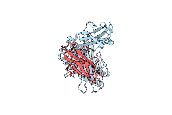 |
Crystal Structure Of Psla-1*0401 Complex With Fmdv-Derived Epitope Mtahitvpy
Organism: Sus scrofa, Foot-and-mouth disease virus
Method: X-RAY DIFFRACTION Resolution:2.50 Å Release Date: 2020-09-09 Classification: STRUCTURAL PROTEIN |
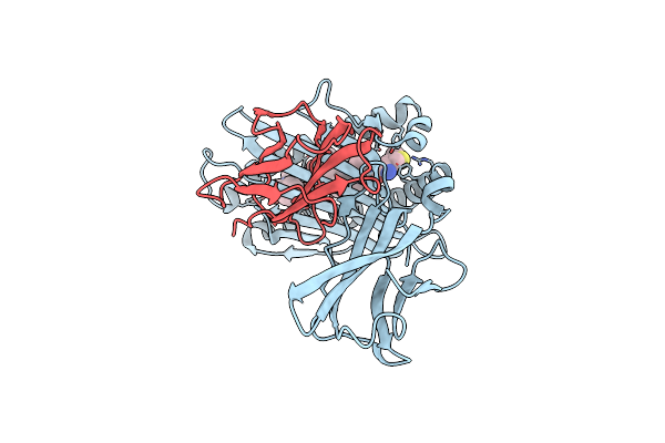 |
Crystal Structure Of Psla-1*0401(R156A) Complex With Fmdv-Derived Epitope Mtahitvpy
Organism: Sus scrofa, Foot-and-mouth disease virus
Method: X-RAY DIFFRACTION Resolution:1.80 Å Release Date: 2020-09-09 Classification: STRUCTURAL PROTEIN |
 |
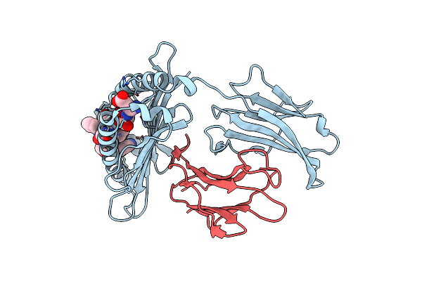
Crystal Structure Of Psla-1*1301(F99Y) Complex With S-Oiv-Derived Epitope Nsdtvgwsw
Organism: Sus scrofa, Swine influenza virus
Method: X-RAY DIFFRACTION Resolution:2.40 Å Release Date: 2020-09-09 Classification: STRUCTURAL PROTEIN |
 |
Organism: Sus scrofa, Neuraminidase deficient flu strains
Method: X-RAY DIFFRACTION Resolution:1.80 Å Release Date: 2020-09-09 Classification: STRUCTURAL PROTEIN |