Search Count: 19
 |
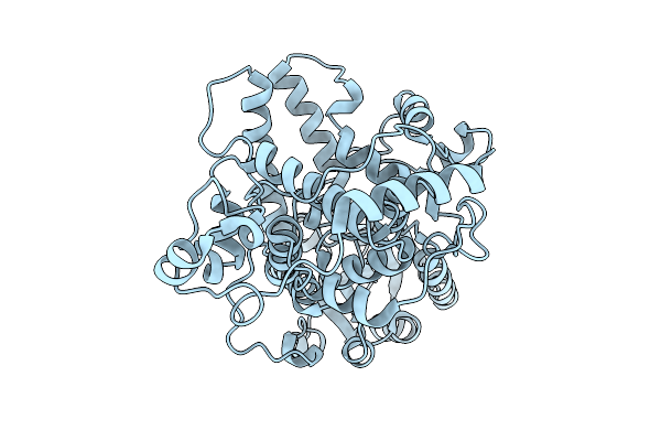
Organism: Erithacus rubecula
Method: X-RAY DIFFRACTION Release Date: 2025-08-27 Classification: CIRCADIAN CLOCK PROTEIN |
 |
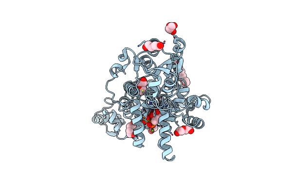
Organism: Arabidopsis thaliana
Method: X-RAY DIFFRACTION Resolution:3.00 Å Release Date: 2021-03-03 Classification: PLANT PROTEIN Ligands: FMN, EDO |
 |
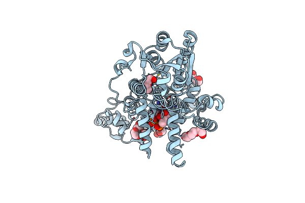
Organism: Arabidopsis thaliana
Method: X-RAY DIFFRACTION Resolution:3.00 Å Release Date: 2021-03-03 Classification: CIRCADIAN CLOCK PROTEIN, PLANT PROTEIN Ligands: FMN, EDO |
 |
Crystal Structure Of Pigeon Cryptochrome 4 Mutant Y319D In Complex With Flavin Adenine Dinucleotide
Organism: Columba livia
Method: X-RAY DIFFRACTION Resolution:1.79 Å Release Date: 2019-09-04 Classification: CIRCADIAN CLOCK PROTEIN Ligands: FAD, MG, PEG, GOL |
 |
Organism: Columba livia
Method: X-RAY DIFFRACTION Resolution:1.90 Å Release Date: 2019-09-04 Classification: CIRCADIAN CLOCK PROTEIN Ligands: FAD, PGE, EDO, PEG, GOL |
 |
2.3 Angstrom Structure Of Phosphodiesterase Treated Vivid (Complex With Fmn)
Organism: Neurospora crassa
Method: X-RAY DIFFRACTION Resolution:2.10 Å Release Date: 2018-03-21 Classification: CIRCADIAN CLOCK PROTEIN Ligands: FMN |
 |
Structure And Kinetics Of The Lov Domain Of Zeitlupe Determine Its Circadian Function In Arabidopsis
Organism: Arabidopsis thaliana
Method: X-RAY DIFFRACTION Resolution:2.50 Å Release Date: 2017-03-08 Classification: CIRCADIAN CLOCK PROTEIN Ligands: FMN |
 |
Structure And Kinetics Of The Lov Domain Of Zeitlupe Determine Its Circadian Function In Arabidopsis
Organism: Arabidopsis thaliana
Method: X-RAY DIFFRACTION Resolution:2.60 Å Release Date: 2017-03-08 Classification: CIRCADIAN CLOCK PROTEIN Ligands: FMN |
 |
Structure And Kinetics Of The Lov Domain Of Zeitlupe Determine Its Circadian Function In Arabidopsis
Organism: Arabidopsis thaliana
Method: X-RAY DIFFRACTION Resolution:2.10 Å Release Date: 2017-03-08 Classification: CIRCADIAN CLOCK PROTEIN Ligands: FMN, ACT, GOL |
 |
Organism: Arabidopsis thaliana
Method: X-RAY DIFFRACTION Resolution:2.29 Å Release Date: 2017-03-08 Classification: CIRCADIAN CLOCK PROTEIN Ligands: FMN |
 |
Structural Biochemistry Of A Fungal Lov Domain Photoreceptor Reveals An Evolutionarily Conserved Pathway Integrating Blue-Light And Oxidative Stress
Organism: Hypocrea jecorina
Method: X-RAY DIFFRACTION Resolution:2.23 Å Release Date: 2015-01-14 Classification: CIRCADIAN CLOCK PROTEIN Ligands: FMN, SO4 |
 |
Organism: Drosophila melanogaster
Method: X-RAY DIFFRACTION Resolution:2.30 Å Release Date: 2012-09-26 Classification: SIGNALING PROTEIN Ligands: MG, FAD |
 |
Organism: Neurospora crassa
Method: X-RAY DIFFRACTION Resolution:2.30 Å Release Date: 2009-11-03 Classification: SIGNALING PROTEIN Ligands: FAD |
 |
Organism: Neurospora crassa
Method: X-RAY DIFFRACTION Resolution:1.80 Å Release Date: 2009-09-29 Classification: SIGNALING PROTEIN Ligands: FAD |
 |
Organism: Neurospora crassa
Method: X-RAY DIFFRACTION Resolution:2.00 Å Release Date: 2009-09-29 Classification: SIGNALING PROTEIN Ligands: FAD |
 |
1.65 Angstrom Crystal Structure Of The Cys71Val Variant In The Fungal Photoreceptor Vvd
Organism: Neurospora crassa
Method: X-RAY DIFFRACTION Resolution:1.65 Å Release Date: 2008-06-17 Classification: SIGNALING PROTEIN Ligands: FAD |
 |
2.0 Angstrom Crystal Structure Of The Fungal Blue-Light Photoreceptor Vivid
Organism: Neurospora crassa
Method: X-RAY DIFFRACTION Resolution:2.00 Å Release Date: 2007-06-05 Classification: CIRCADIAN CLOCK PROTEIN Ligands: FAD |
 |
Organism: Neurospora crassa
Method: X-RAY DIFFRACTION Resolution:1.80 Å Release Date: 2007-06-05 Classification: CIRCADIAN CLOCK PROTEIN Ligands: FAD |
 |
1.7 Angstrom Crystal Structure Of The Photo-Excited Blue-Light Photoreceptor Vivid
Organism: Neurospora crassa
Method: X-RAY DIFFRACTION Resolution:1.70 Å Release Date: 2007-06-05 Classification: CIRCADIAN CLOCK PROTEIN Ligands: FAD |

