
Search Count: 29
 |
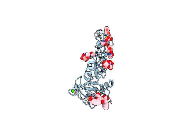
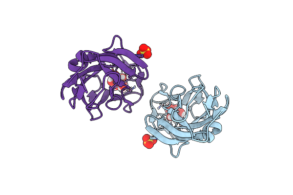
Crystal Structure Of Fimh In Complex With A Tetraflourinated Biphenyl Alpha D-Mannoside
Organism: Escherichia coli (strain k12)
Method: X-RAY DIFFRACTION Resolution:2.10 Å Release Date: 2019-03-20 Classification: CELL ADHESION Ligands: EJK, SO4, EPE |
 |
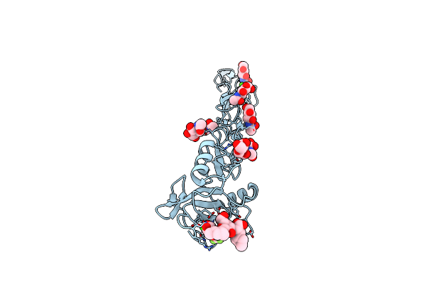
Crystal Structure Of Fimh In Complex With A Pentaflourinated Biphenyl Alpha D-Mannoside
Organism: Escherichia coli (strain k12)
Method: X-RAY DIFFRACTION Resolution:2.20 Å Release Date: 2019-03-20 Classification: CELL ADHESION Ligands: EJN, SO4 |
 |
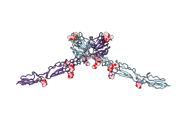
E-Selectin Lectin, Egf-Like And Two Scr Domains Complexed With Glycomimetic Ligand Bw69669
Organism: Homo sapiens
Method: X-RAY DIFFRACTION Resolution:2.04 Å Release Date: 2018-11-21 Classification: CELL ADHESION Ligands: NAG, C4Z, CA |
 |
E-Selectin Lectin, Egf-Like And Two Scr Domains Complexed With Glycomimetic Ligand Nv354
Organism: Homo sapiens
Method: X-RAY DIFFRACTION Resolution:2.20 Å Release Date: 2018-11-21 Classification: CELL ADHESION Ligands: NAG, C5H, CA |
 |
E-Selectin Lectin, Egf-Like And Two Scr Domains Complexed With Glycomimetic Ligand Nv355
Organism: Homo sapiens
Method: X-RAY DIFFRACTION Resolution:2.21 Å Release Date: 2018-11-21 Classification: CELL ADHESION Ligands: NAG, C5K, CA |
 |
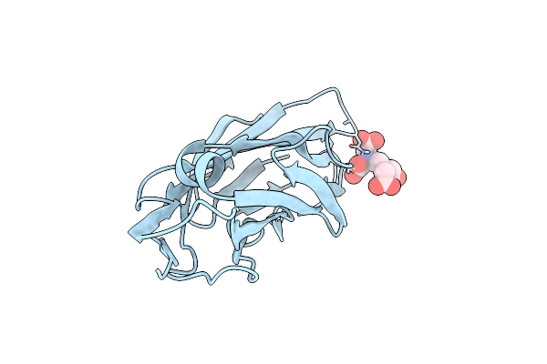
Crystal Structure Of The Fimh Lectin Domain In Complex With 1,5-Anhydromannitol
Organism: Escherichia coli (strain k12)
Method: X-RAY DIFFRACTION Resolution:2.60 Å Release Date: 2018-02-14 Classification: SUGAR BINDING PROTEIN Ligands: AH2 |
 |
Crystal Structure Of Fimh Lectin Domain In Complex With 2-Deoxy-Heptylmannoside
Organism: Escherichia coli k12, Escherichia coli k-12
Method: X-RAY DIFFRACTION Resolution:1.90 Å Release Date: 2017-06-21 Classification: SUGAR BINDING PROTEIN Ligands: 6LS |
 |
Crystal Structure Of Fimh Lectin Domain In Complex With 2-Fluoro-Heptylmannoside
Organism: Escherichia coli (strain k12)
Method: X-RAY DIFFRACTION Resolution:2.10 Å Release Date: 2017-06-21 Classification: SUGAR BINDING PROTEIN Ligands: 6KS |
 |
Crystal Structure Of Fimh Lectin Domain In Complex With 3-Deoxy-Heptylmannoside
Organism: Escherichia coli (strain k12)
Method: X-RAY DIFFRACTION Resolution:2.99 Å Release Date: 2017-06-21 Classification: SUGAR BINDING PROTEIN Ligands: 6KW |
 |
Crystal Structure Of Fimh Lectin Domain In Complex With 3-Fluoro-Heptylmannoside
Organism: Escherichia coli (strain k12)
Method: X-RAY DIFFRACTION Resolution:1.90 Å Release Date: 2017-06-21 Classification: SUGAR BINDING PROTEIN Ligands: 6KH |
 |
Crystal Structure Of Fimh Lectin Domain In Complex With 4-Deoxy-Heptylmannoside
Organism: Escherichia coli (strain k12)
Method: X-RAY DIFFRACTION Resolution:1.90 Å Release Date: 2017-06-21 Classification: SUGAR BINDING PROTEIN Ligands: 6KU |
 |
Crystal Structure Of Fimh Lectin Domain In Complex With 4-Fluoro-Heptylmannoside
Organism: Escherichia coli (strain k12)
Method: X-RAY DIFFRACTION Resolution:1.90 Å Release Date: 2017-06-21 Classification: SUGAR BINDING PROTEIN Ligands: 6K3 |
 |
Breaking Down The Wall: Mutation Of The Tyrosine Gate Of The Universal Escherichia Coli Fimbrial Adhesin Fimh
Organism: Escherichia coli
Method: X-RAY DIFFRACTION Resolution:1.90 Å Release Date: 2017-01-18 Classification: CELL ADHESION Ligands: EDT |
 |
Breaking Down The Wall: Mutation Of The Tyrosine Gate Of The Universal Escherichia Coli Fimbrial Adhesin Fimh
Organism: Escherichia coli
Method: X-RAY DIFFRACTION Resolution:1.42 Å Release Date: 2016-11-30 Classification: CELL ADHESION Ligands: KGM, NA |
 |
Breaking Down The Wall: Mutation Of The Tyrosine Gate Of The Universal Escherichia Coli Fimbrial Adhesin Fimh
Organism: Escherichia coli
Method: X-RAY DIFFRACTION Resolution:2.13 Å Release Date: 2016-11-30 Classification: CELL ADHESION Ligands: 3X8 |
 |
Organism: Escherichia coli k-12
Method: X-RAY DIFFRACTION Resolution:1.60 Å Release Date: 2016-07-20 Classification: SUGAR BINDING PROTEIN Ligands: 51C, SO4 |
 |
Crystal Structure Of The Lectin Domain Of Papg From E. Coli Bi47 In Complex With Ssea4 In Space Group P212121
Organism: Escherichia coli
Method: X-RAY DIFFRACTION Resolution:1.80 Å Release Date: 2016-04-13 Classification: SUGAR BINDING PROTEIN |
 |
Crystal Structure Of The Lectin Domain Of Papg From E. Coli Bi47 In Complex With Ssea4 In Space Group P21
Organism: Escherichia coli
Method: X-RAY DIFFRACTION Resolution:1.80 Å Release Date: 2016-04-13 Classification: SUGAR BINDING PROTEIN Ligands: ZN |
 |
Crystal Structure Of The Lectin Domain Of Papg From E. Coli Bi47 In Complex With 4-Methoxyphenyl Beta-D-Galabiose In Space Group P212121
Organism: Escherichia coli
Method: X-RAY DIFFRACTION Resolution:1.45 Å Release Date: 2016-04-13 Classification: SUGAR BINDING PROTEIN Ligands: MES, 4KS |
 |
Crystal Structure Of The Lectin Domain Of Papg From E. Coli Bi47 In Complex With 4-Methoxyphenyl Beta-D-Galabiose In Space Group P21
Organism: Escherichia coli
Method: X-RAY DIFFRACTION Resolution:1.50 Å Release Date: 2016-04-13 Classification: SUGAR BINDING PROTEIN Ligands: 4KS |