
Search Count: 17
 |
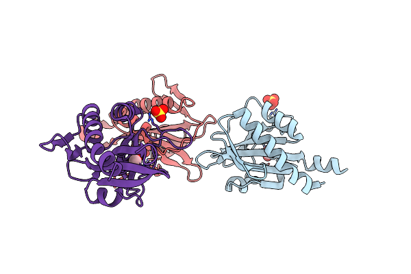
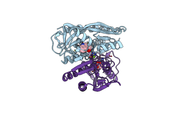
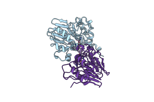
Ligand Binding Domain Of Roseburia Intestinalis L1-82 Uracil Chemoreceptor (Dcache) In Complex With Uracil And Acetate
Organism: Roseburia intestinalis l1-82
Method: X-RAY DIFFRACTION Resolution:1.46 Å Release Date: 2025-03-12 Classification: SIGNALING PROTEIN Ligands: URA, ACT, EDO |
 |
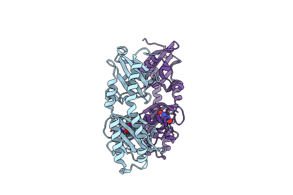
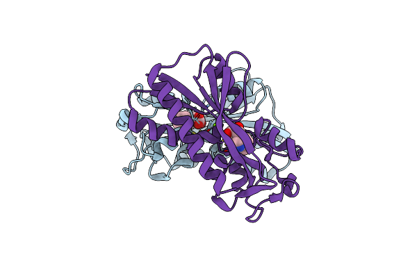
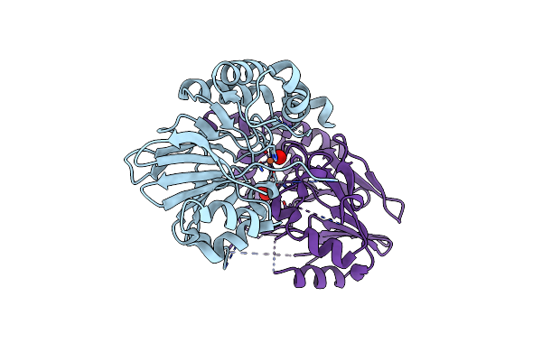
Sensor Domain Of Oscillibacter Ruminantium Chemoreceptor In Complex With Formate.
Organism: Oscillibacter ruminantium
Method: X-RAY DIFFRACTION Resolution:1.75 Å Release Date: 2025-02-12 Classification: SIGNALING PROTEIN Ligands: FMT, NA |
 |
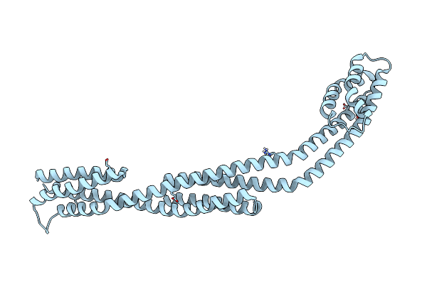
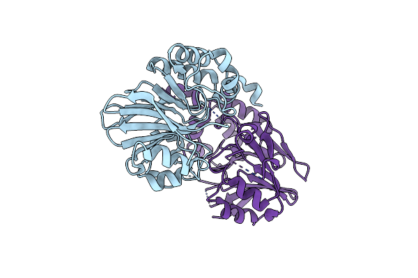
Sensor Domain Of Asticcacaulis Benevestitus Chemoreceptor In Complex With Formate.
Organism: Asticcacaulis benevestitus
Method: X-RAY DIFFRACTION Resolution:2.10 Å Release Date: 2025-02-12 Classification: SIGNALING PROTEIN Ligands: FMT, SO4 |
 |
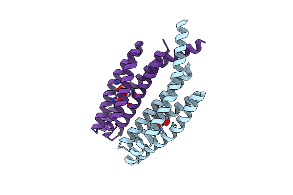
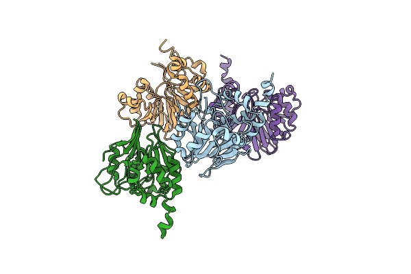
Ligand Binding Domain Of The P. Putida Receptor Mcph In Complex With Uric Acid
Organism: Pseudomonas putida kt2440
Method: X-RAY DIFFRACTION Resolution:1.95 Å Release Date: 2024-07-24 Classification: SIGNALING PROTEIN Ligands: URC |
 |
Organism: Sinorhizobium meliloti
Method: X-RAY DIFFRACTION Resolution:2.70 Å Release Date: 2023-07-05 Classification: SIGNALING PROTEIN Ligands: YB |
 |
Organism: Comamonas testosteroni cnb-2
Method: X-RAY DIFFRACTION Resolution:1.80 Å Release Date: 2023-01-04 Classification: SIGNALING PROTEIN Ligands: LMR |
 |
Structure Of The Ligand Binding Domain Of The Antibiotic Biosynthesis Regulator Admx From The Rhizobacterium Serratia Plymuthica A153 Bound To The Auxin Indole-3-Acetic Acid (Iaa).
Organism: Serratia plymuthica
Method: X-RAY DIFFRACTION Resolution:1.81 Å Release Date: 2022-12-14 Classification: TRANSCRIPTION Ligands: IAC, MG |
 |
Structure Of The Ligand Binding Domain Of The Antibiotic Biosynthesis Regulator Admx From The Rhizobacterium Serratia Plymuthica A153 Bound To The Auxin Indole-3-Piruvic Acid (Ipa).
Organism: Serratia plymuthica
Method: X-RAY DIFFRACTION Resolution:2.25 Å Release Date: 2022-12-14 Classification: TRANSCRIPTION Ligands: 3IO, MG |
 |
Organism: Treponema denticola atcc 35404
Method: X-RAY DIFFRACTION Resolution:2.10 Å Release Date: 2019-06-19 Classification: SIGNALING PROTEIN |
 |
Organism: Thermotoga maritima
Method: X-RAY DIFFRACTION Resolution:2.56 Å Release Date: 2019-06-19 Classification: SIGNALING PROTEIN |
 |
Organism: Thermotoga maritima
Method: X-RAY DIFFRACTION Resolution:2.00 Å Release Date: 2019-06-19 Classification: SIGNALING PROTEIN Ligands: ZN |
 |
Organism: Treponema denticola atcc 35404
Method: X-RAY DIFFRACTION Resolution:2.07 Å Release Date: 2019-06-19 Classification: SIGNALING PROTEIN Ligands: FE, PER, O, CL |
 |
Organism: Comamonas testosteroni (strain cnb-2)
Method: X-RAY DIFFRACTION Resolution:2.50 Å Release Date: 2018-12-26 Classification: SIGNALING PROTEIN Ligands: CIT |
 |
Organism: Comamonas testosteroni
Method: X-RAY DIFFRACTION Resolution:2.80 Å Release Date: 2018-06-27 Classification: SIGNALING PROTEIN |
 |
Organism: Comamonas testosteroni (strain cnb-2)
Method: X-RAY DIFFRACTION Resolution:2.50 Å Release Date: 2018-06-27 Classification: SIGNALING PROTEIN Ligands: CIT |
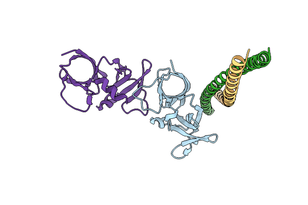 |
The Structure Of A Ternary Complex Between Chea Domains P4 And P5 With Chew And With An Unzipped Fragment Of Tm14, A Chemoreceptor Analog From Thermotoga Maritima.
Organism: Thermotoga maritima
Method: X-RAY DIFFRACTION Resolution:3.19 Å Release Date: 2013-08-28 Classification: IMMUNE SYSTEM |
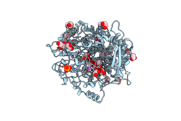 |
Organism: Hahella chejuensis
Method: X-RAY DIFFRACTION Resolution:1.75 Å Release Date: 2012-10-17 Classification: HYDROLASE Ligands: PEG, GOL, PO4, ACT, EDO, CA, NA |