
Search Count: 300
 |
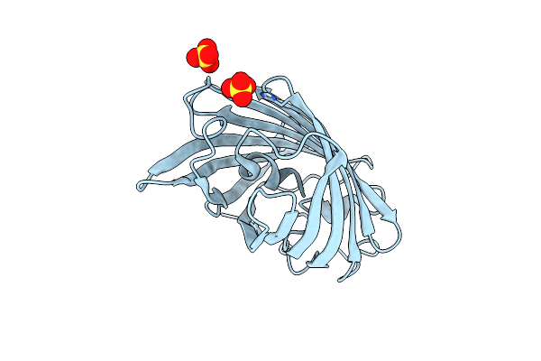
Organism: Caldicellulosiruptor sp.
Method: X-RAY DIFFRACTION Release Date: 2025-10-15 Classification: HYDROLASE Ligands: GOL, SO4 |
 |
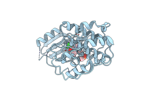
Crystal Structure Of Beta-Glucosidase Cabgl Mutant E163Q In Complex With Glucose
Organism: Caldicellulosiruptor sp.
Method: X-RAY DIFFRACTION Release Date: 2025-10-15 Classification: HYDROLASE Ligands: GOL, BGC, SO4 |
 |
Organism: Homo sapiens
Method: ELECTRON MICROSCOPY Release Date: 2025-10-01 Classification: EXOCYTOSIS Ligands: GNP |
 |
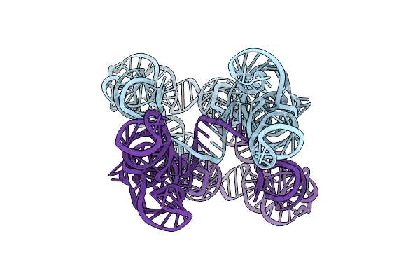
Organism: Homo sapiens, Synthetic construct
Method: ELECTRON MICROSCOPY Release Date: 2025-08-13 Classification: STRUCTURAL PROTEIN/DNA |
 |
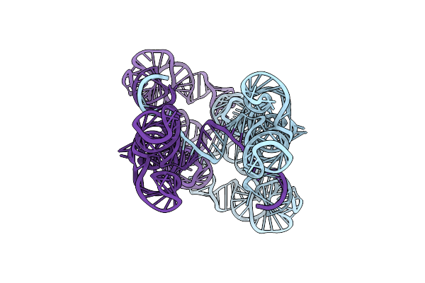
Organism: Homo sapiens, Synthetic construct
Method: ELECTRON MICROSCOPY Release Date: 2025-08-13 Classification: STRUCTURAL PROTEIN/DNA |
 |
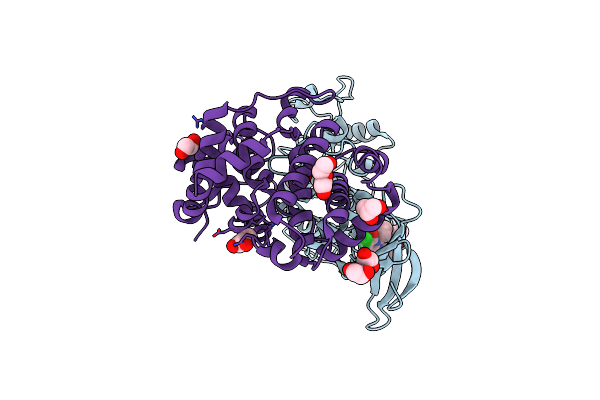
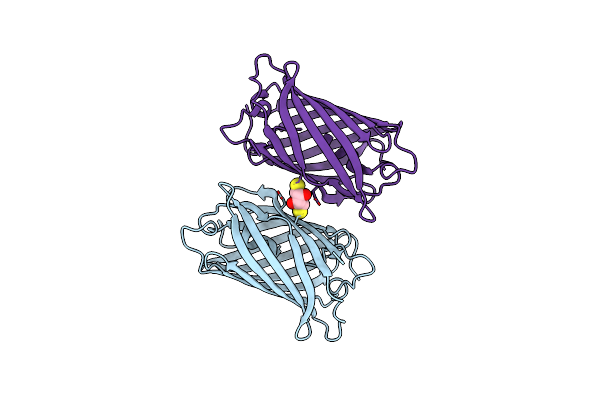
Organism: Homo sapiens
Method: X-RAY DIFFRACTION Resolution:2.60 Å Release Date: 2025-04-16 Classification: CELL CYCLE Ligands: A1A1H |
 |
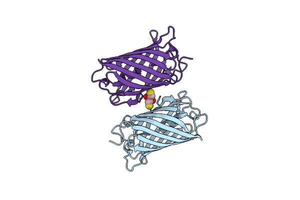
Organism: Homo sapiens
Method: X-RAY DIFFRACTION Resolution:2.54 Å Release Date: 2025-04-16 Classification: CELL CYCLE Ligands: A1A1H, PEG, GOL, EDO |
 |
Organism: Homo sapiens
Method: ELECTRON MICROSCOPY Release Date: 2025-04-16 Classification: CELL CYCLE Ligands: ZN, A1A1I |
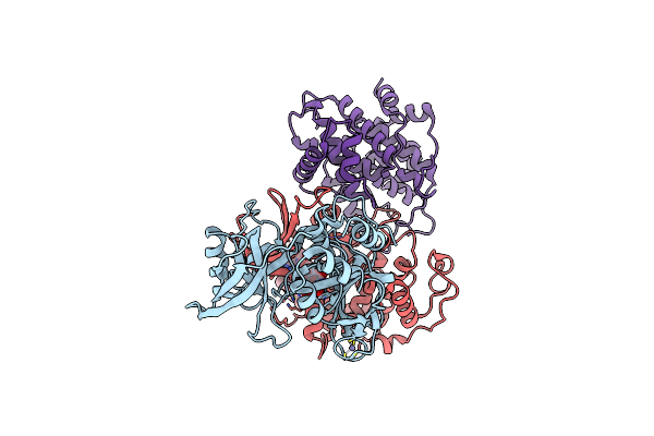 |
Cryo-Em Structure Of Cdk2/Cycline1 In Complex With Crbn/Ddb1 And Cpd 4 (Local Mask)
Organism: Homo sapiens
Method: ELECTRON MICROSCOPY Release Date: 2025-04-16 Classification: CELL CYCLE Ligands: ZN, A1A1I |
 |
Organism: Lobophyllia hemprichii
Method: X-RAY DIFFRACTION Resolution:1.34 Å Release Date: 2025-04-16 Classification: FLUORESCENT PROTEIN Ligands: SO4 |
 |
Organism: Lobophyllia hemprichii
Method: X-RAY DIFFRACTION Resolution:1.66 Å Release Date: 2025-04-16 Classification: FLUORESCENT PROTEIN Ligands: DTT |
 |
Organism: Lobophyllia hemprichii
Method: X-RAY DIFFRACTION Resolution:1.85 Å Release Date: 2025-04-16 Classification: FLUORESCENT PROTEIN Ligands: DTT |
 |
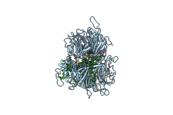
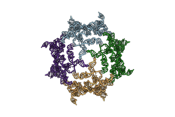
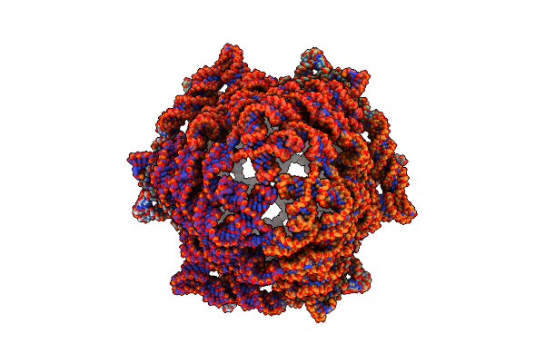
Organism: Enterococcus faecalis
Method: ELECTRON MICROSCOPY Release Date: 2025-03-19 Classification: RNA |
 |
Organism: Enterococcus faecalis jh1
Method: ELECTRON MICROSCOPY Release Date: 2025-03-19 Classification: RNA |
 |
Organism: Enterococcus faecalis
Method: ELECTRON MICROSCOPY Release Date: 2025-03-19 Classification: RNA |
 |
Organism: Ligilactobacillus salivarius dsm 20555 = atcc 11741
Method: ELECTRON MICROSCOPY Release Date: 2025-03-19 Classification: RNA Ligands: MG |
 |
Organism: Fusobacterium nucleatum
Method: ELECTRON MICROSCOPY Release Date: 2025-03-19 Classification: RNA |
 |
Organism: Fusobacterium nucleatum
Method: ELECTRON MICROSCOPY Release Date: 2025-03-19 Classification: RNA |
 |
Organism: Streptococcus agalactiae
Method: ELECTRON MICROSCOPY Release Date: 2025-03-19 Classification: RNA |
 |
Organism: Clostridium botulinum a str. atcc 19397
Method: ELECTRON MICROSCOPY Resolution:2.60 Å Release Date: 2025-03-19 Classification: RNA Ligands: MG |