
Search Count: 158
 |
Organism: Escherichia coli (strain k12), Bacteroides fragilis
Method: X-RAY DIFFRACTION Release Date: 2025-10-01 Classification: TOXIN |
 |
Organism: Bacteroides fragilis
Method: X-RAY DIFFRACTION Release Date: 2025-10-01 Classification: ANTITOXIN Ligands: IPA |
 |
Organism: Bacteroides fragilis nctc 9343
Method: X-RAY DIFFRACTION Release Date: 2025-10-01 Classification: STRUCTURAL PROTEIN |
 |
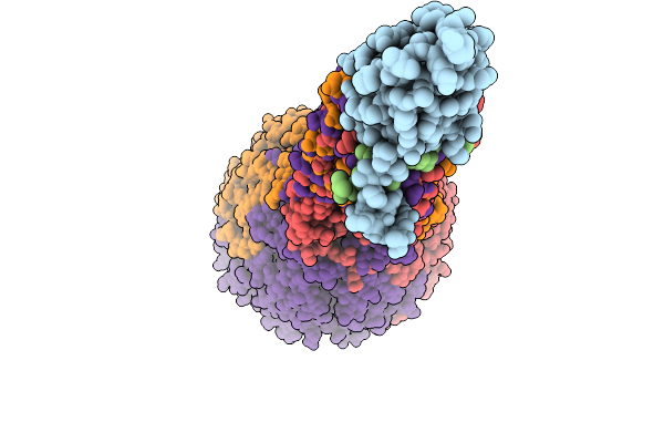
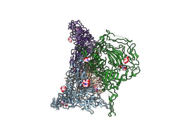
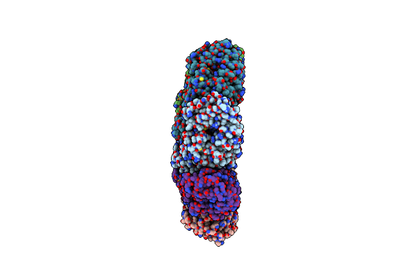
Structure Of Cargo Complex (Btpea-Btaeb-Btapc) Bound To The Vgrg Spike From The Type Vi Secretion System
Organism: Bacteroides fragilis
Method: ELECTRON MICROSCOPY Release Date: 2025-09-10 Classification: ANTIMICROBIAL PROTEIN Ligands: ZN |
 |
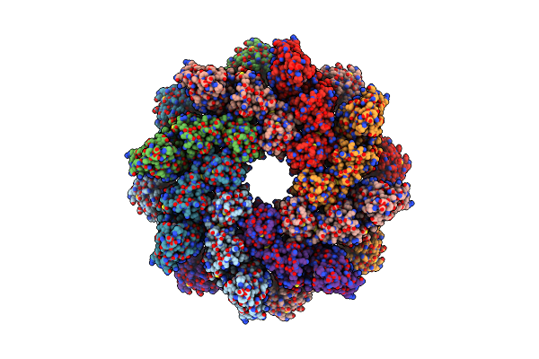
Structure Of The Btpea Effector And Btaeb Effector Bound To The Vgrg Spike From The Type Vi Secretion System
Organism: Bacteroides fragilis
Method: ELECTRON MICROSCOPY Release Date: 2025-09-10 Classification: ANTIMICROBIAL PROTEIN Ligands: ZN |
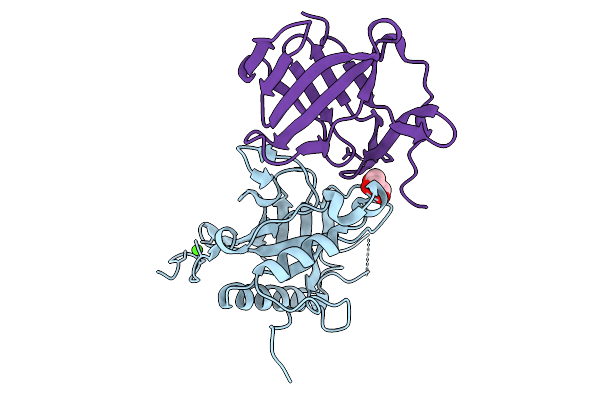 |
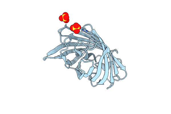
Crystal Structure Of The Type Vi Secretion System Effector-Immunity Complex Btaeb Ctd-Btaib From Bacteroides Fragilis
Organism: Bacteroides fragilis
Method: X-RAY DIFFRACTION Release Date: 2025-09-03 Classification: ANTIMICROBIAL PROTEIN Ligands: GOL, CA |
 |
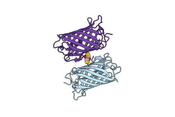
Organism: Bacteroides fragilis
Method: X-RAY DIFFRACTION Release Date: 2025-09-03 Classification: ANTIMICROBIAL PROTEIN Ligands: PEG |
 |
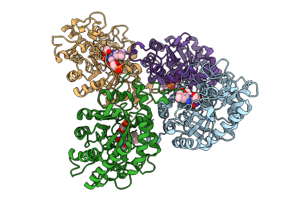
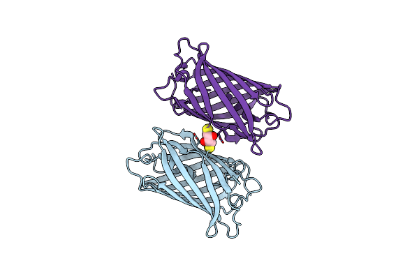
Crystal Structure Of The Type Vi Secretion System Effector-Immunity Complex Btpea Ctd-Btpia From Bacteroides Fragilis.
Organism: Bacteroides fragilis
Method: X-RAY DIFFRACTION Release Date: 2025-09-03 Classification: ANTIMICROBIAL PROTEIN Ligands: GOL |
 |
Organism: Parageobacillus thermantarcticus
Method: X-RAY DIFFRACTION Release Date: 2025-08-27 Classification: OXIDOREDUCTASE Ligands: ACT, FMN |
 |
Organism: Mus musculus
Method: ELECTRON MICROSCOPY Release Date: 2025-07-23 Classification: MEMBRANE PROTEIN Ligands: NAG |
 |
Organism: Mus musculus
Method: ELECTRON MICROSCOPY Release Date: 2025-07-23 Classification: MEMBRANE PROTEIN Ligands: NAG, CU |
 |
Organism: Lobophyllia hemprichii
Method: X-RAY DIFFRACTION Resolution:1.34 Å Release Date: 2025-04-16 Classification: FLUORESCENT PROTEIN Ligands: SO4 |
 |
Organism: Lobophyllia hemprichii
Method: X-RAY DIFFRACTION Resolution:1.66 Å Release Date: 2025-04-16 Classification: FLUORESCENT PROTEIN Ligands: DTT |
 |
Organism: Lobophyllia hemprichii
Method: X-RAY DIFFRACTION Resolution:1.85 Å Release Date: 2025-04-16 Classification: FLUORESCENT PROTEIN Ligands: DTT |
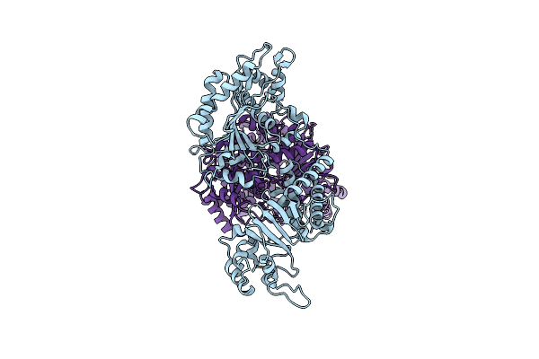 |
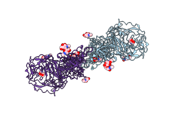
Cryo-Electron Microscopy (Cryo-Em) Structure Of The Hachiman Defense System From Escherichia Coli
Organism: Escherichia coli k-12
Method: ELECTRON MICROSCOPY Release Date: 2025-03-26 Classification: DNA BINDING PROTEIN |
 |
Organism: Homo sapiens
Method: ELECTRON MICROSCOPY Release Date: 2025-03-19 Classification: HYDROLASE Ligands: A1EIV |
 |
Organism: Homo sapiens
Method: ELECTRON MICROSCOPY Release Date: 2025-03-19 Classification: HYDROLASE Ligands: A1EIV |
 |
Organism: Homo sapiens
Method: ELECTRON MICROSCOPY Release Date: 2025-03-12 Classification: HYDROLASE Ligands: A1EIV |
 |
Organism: Homo sapiens
Method: ELECTRON MICROSCOPY Release Date: 2025-03-12 Classification: HYDROLASE |
 |
Organism: Arabidopsis thaliana
Method: ELECTRON MICROSCOPY Release Date: 2025-03-05 Classification: TRANSPORT PROTEIN |