
Search Count: 351
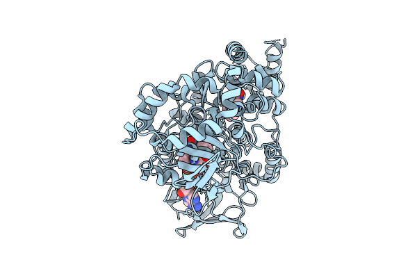 |
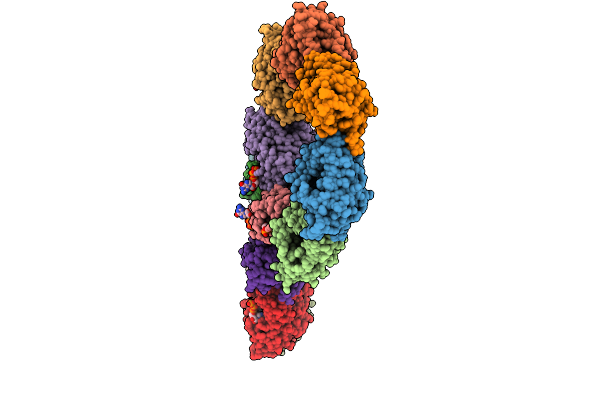
Organism: Aetokthonos hydrillicola thurmond2011
Method: X-RAY DIFFRACTION Release Date: 2025-12-10 Classification: FLAVOPROTEIN Ligands: FAD, TRP |
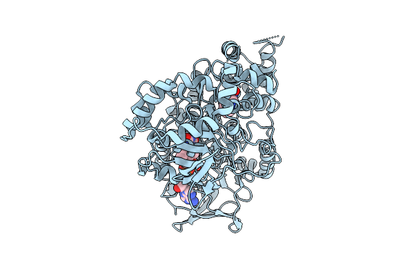 |
Organism: Aetokthonos hydrillicola thurmond2011
Method: X-RAY DIFFRACTION Release Date: 2025-12-10 Classification: FLAVOPROTEIN Ligands: FAD, TRP |
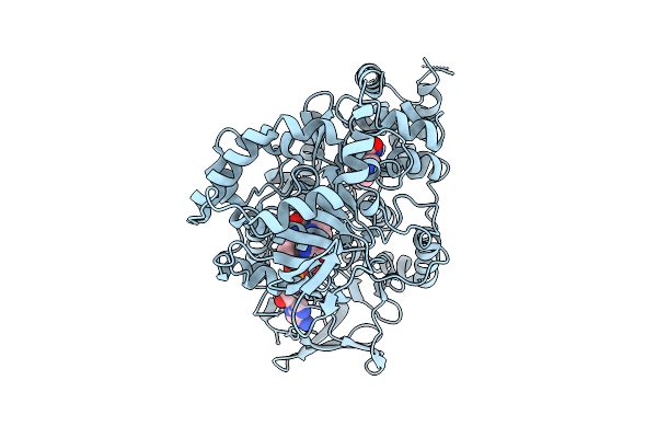 |
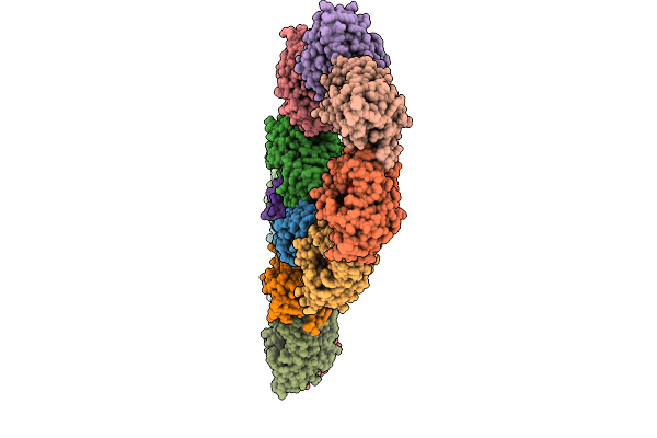
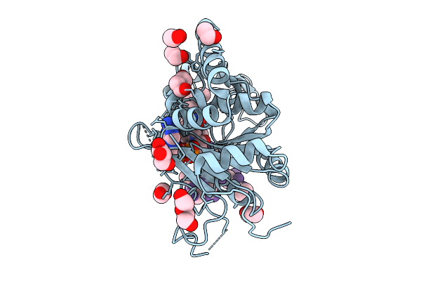
Crystal Structure Of Aetf-L183F/V220I/S523A In Complex With Fad And L-Tryptophan
Organism: Aetokthonos hydrillicola thurmond2011
Method: X-RAY DIFFRACTION Release Date: 2025-12-10 Classification: FLAVOPROTEIN Ligands: TRP, FAD |
 |
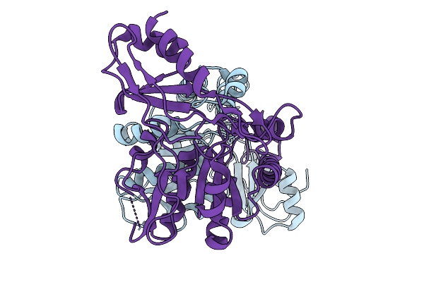
Organism: Homo sapiens
Method: X-RAY DIFFRACTION Release Date: 2025-11-05 Classification: IMMUNE SYSTEM |
 |
Organism: Homo sapiens
Method: X-RAY DIFFRACTION Release Date: 2025-11-05 Classification: IMMUNE SYSTEM Ligands: PO4 |
 |
Organism: Erwinia amylovora atcc 49946
Method: X-RAY DIFFRACTION Release Date: 2025-10-01 Classification: LYASE |
 |
Organism: Erwinia amylovora atcc 49946
Method: X-RAY DIFFRACTION Release Date: 2025-10-01 Classification: BIOSYNTHETIC PROTEIN |
 |
Organism: Erwinia amylovora atcc 49946
Method: X-RAY DIFFRACTION Release Date: 2025-10-01 Classification: BIOSYNTHETIC PROTEIN Ligands: ATP |
 |
Organism: Erwinia amylovora atcc 49946
Method: X-RAY DIFFRACTION Release Date: 2025-10-01 Classification: BIOSYNTHETIC PROTEIN Ligands: GTP |
 |
Organism: Erwinia amylovora atcc 49946
Method: X-RAY DIFFRACTION Release Date: 2025-10-01 Classification: BIOSYNTHETIC PROTEIN Ligands: ATP, GTP, MG |
 |
Sirt2 Structure In Complex With H3K18Myr Peptide And Native Nad: Pre-Catalysis State 3
Organism: Homo sapiens
Method: X-RAY DIFFRACTION Release Date: 2025-09-24 Classification: HYDROLASE Ligands: PEG, EDO, NAD, GOL, ZN, MYR |
 |
Sirt2 Structure In Complex With H3K18Myr Peptide And Native Nad: Pre-Catalysis State 1
Organism: Homo sapiens
Method: X-RAY DIFFRACTION Release Date: 2025-09-24 Classification: HYDROLASE Ligands: NAD, PEG, EDO, GOL, ZN, MYR |
 |
Sirt2 Structure In Complex With H3K18Myr Peptide And Native Nad: Pre-Catalysis State 2
Organism: Homo sapiens
Method: X-RAY DIFFRACTION Release Date: 2025-09-24 Classification: HYDROLASE Ligands: NAD, EDO, GOL, ZN, MYR |
 |
Organism: Homo sapiens
Method: X-RAY DIFFRACTION Release Date: 2025-09-24 Classification: HYDROLASE Ligands: EDO, ZN, MYR |
 |
Organism: Homo sapiens
Method: X-RAY DIFFRACTION Release Date: 2025-09-24 Classification: HYDROLASE Ligands: EDO, ZN, MYR |
 |
Organism: Homo sapiens
Method: X-RAY DIFFRACTION Release Date: 2025-09-24 Classification: HYDROLASE Ligands: NCA, GOL, ZN, YDD |
 |
Organism: Homo sapiens
Method: X-RAY DIFFRACTION Release Date: 2025-09-24 Classification: HYDROLASE Ligands: NAD, EDO, PEG, ZN, MYR |
 |
Organism: Homo sapiens
Method: X-RAY DIFFRACTION Release Date: 2025-09-24 Classification: HYDROLASE Ligands: NAD, ZN, MYR |
 |
Crystal Structure Of Sars-Cov-2 Rbd In Complex With A Neutralizing Antibody Scfv N1
Organism: Homo sapiens, Severe acute respiratory syndrome coronavirus 2
Method: X-RAY DIFFRACTION Release Date: 2025-09-03 Classification: VIRAL PROTEIN/IMMUNE SYSTEM Ligands: NAG |
 |
The Glycoprotein E Of Varicella-Zoster Virus In Complex With Wll-1/Wll-28 Fab
Organism: Varicella-zoster virus (strain oka vaccine), Homo sapiens
Method: ELECTRON MICROSCOPY Release Date: 2025-09-03 Classification: VIRAL PROTEIN/IMMUNE SYSTEM |