
Search Count: 22
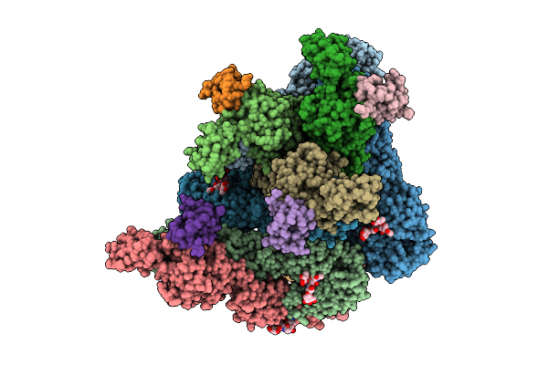 |
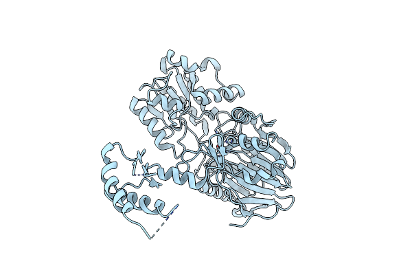
Organism: Chikungunya virus
Method: ELECTRON MICROSCOPY Release Date: 2025-12-03 Classification: VIRUS |
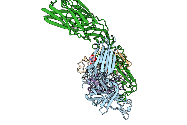 |
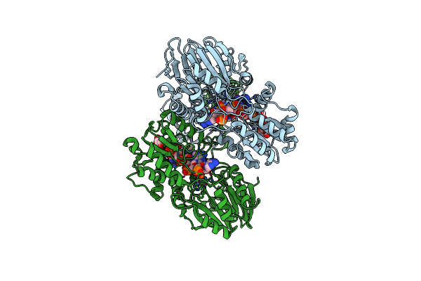
Organism: Chikungunya virus, Homo sapiens
Method: ELECTRON MICROSCOPY Release Date: 2025-11-19 Classification: ANTIFUNGAL PROTEIN Ligands: NAG |
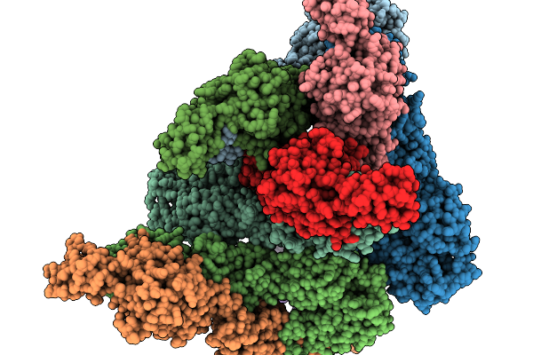 |
Organism: Chikungunya virus
Method: ELECTRON MICROSCOPY Release Date: 2025-11-19 Classification: VIRUS LIKE PARTICLE |
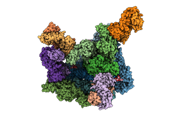 |
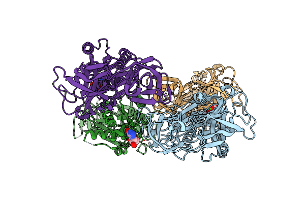
Organism: Homo sapiens, Chikungunya virus
Method: ELECTRON MICROSCOPY Release Date: 2025-11-12 Classification: VIRUS LIKE PARTICLE Ligands: CLR |
 |
Organism: Chikungunya virus, Homo sapiens
Method: X-RAY DIFFRACTION Release Date: 2025-07-30 Classification: ANTIVIRAL PROTEIN |
 |
Organism: Mycobacterium tuberculosis h37rv
Method: X-RAY DIFFRACTION Resolution:2.44 Å Release Date: 2023-01-25 Classification: RNA BINDING PROTEIN Ligands: ZN |
 |
Organism: Mycobacterium tuberculosis h37rv, Synthetic construct
Method: X-RAY DIFFRACTION Resolution:3.20 Å Release Date: 2023-01-25 Classification: RNA BINDING PROTEIN/RNA Ligands: ZN |
 |
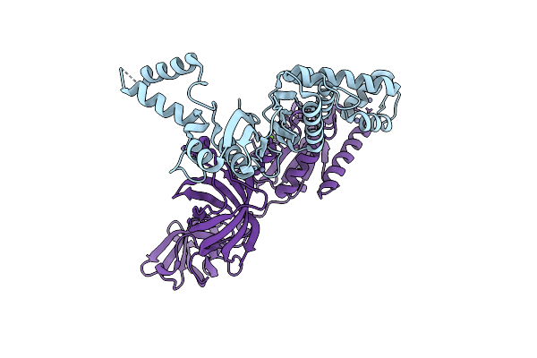
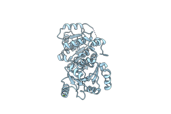
Organism: Mycobacterium tuberculosis
Method: X-RAY DIFFRACTION Resolution:3.40 Å Release Date: 2022-10-19 Classification: Elongation factor Ligands: GDP, MG |
 |
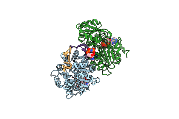
The Crystal Structure Of Ef-Tu And Ef-Ts Complex From Mycobacterium Tuberculosis
Organism: Mycobacterium tuberculosis
Method: X-RAY DIFFRACTION Resolution:2.80 Å Release Date: 2022-10-12 Classification: Elongation Factor Ligands: ZN, MG |
 |
Organism: Homo sapiens
Method: X-RAY DIFFRACTION Resolution:3.10 Å Release Date: 2020-06-17 Classification: RNA BINDING PROTEIN Ligands: MG |
 |
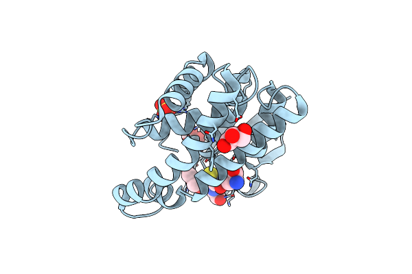
Organism: Homo sapiens
Method: X-RAY DIFFRACTION Resolution:2.70 Å Release Date: 2020-06-17 Classification: RNA BINDING PROTEIN Ligands: AMP |
 |
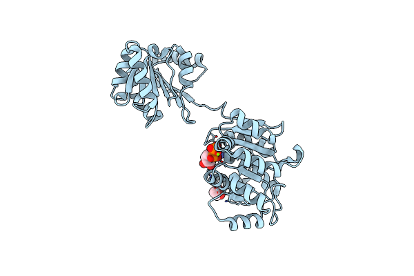
Crystal Structure Of Human Dead-Box Rna Helicase Ddx21 At Post-Unwound State
Organism: Homo sapiens
Method: X-RAY DIFFRACTION Resolution:2.24 Å Release Date: 2020-06-17 Classification: RNA BINDING PROTEIN/RNA Ligands: ANP, MG |
 |
Crystal Structure Of Human Dead-Box Rna Helicase Ddx21 At Post-Hydrolysis State
Organism: Homo sapiens
Method: X-RAY DIFFRACTION Resolution:1.80 Å Release Date: 2020-06-17 Classification: RNA BINDING PROTEIN Ligands: ADP, MG, GOL |
 |
Organism: Mycobacterium tuberculosis (strain atcc 25618 / h37rv)
Method: X-RAY DIFFRACTION Resolution:1.80 Å Release Date: 2019-03-20 Classification: RIBOSOMAL PROTEIN Ligands: GOL, EDO |
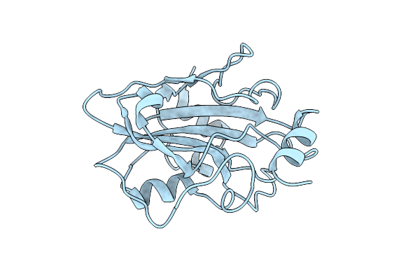 |
Organism: Trichuris muris
Method: X-RAY DIFFRACTION Resolution:1.73 Å Release Date: 2017-08-30 Classification: UNKNOWN FUNCTION |
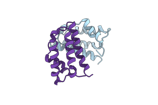 |
Organism: Lutzomyia longipalpis
Method: X-RAY DIFFRACTION Resolution:1.94 Å Release Date: 2016-07-27 Classification: odorant-binding protein |
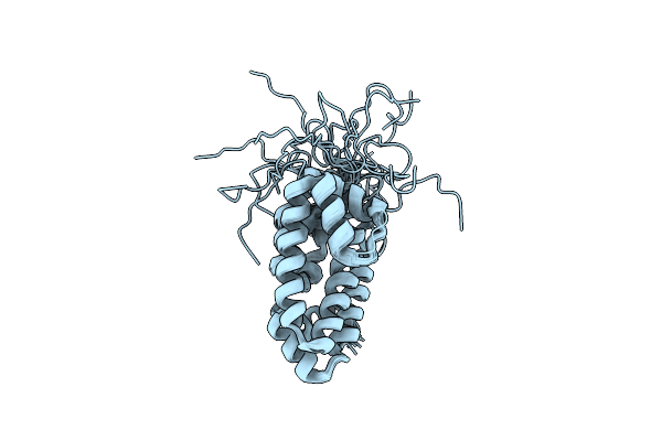 |
Diversity In The Structures And Ligand Binding Sites Among The Fatty Acid And Retinol Binding Proteins Of Nematodes Revealed By Na-Far-1 From Necator Americanus
Organism: Necator americanus
Method: SOLUTION NMR Release Date: 2015-09-16 Classification: RETINOL-BINDING PROTEIN |
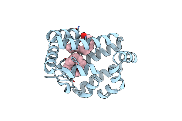 |
Organism: Necator americanus
Method: X-RAY DIFFRACTION Resolution:2.14 Å Release Date: 2015-09-16 Classification: retinol-binding protein Ligands: PLM |
 |
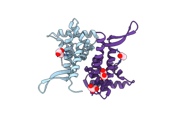
Crystal Structure Of Monomeric Na-Gst-3, A Glutathione S-Transferase From The Major Human Hookworm Parasite Necator Americanus
Organism: Necator americanus
Method: X-RAY DIFFRACTION Resolution:2.07 Å Release Date: 2013-08-14 Classification: TRANSFERASE Ligands: GSH, SO4, GOL |
 |
Organism: Necator americanus
Method: X-RAY DIFFRACTION Resolution:1.90 Å Release Date: 2007-08-07 Classification: TRANSFERASE Ligands: EDO, GSH |