
Search Count: 49
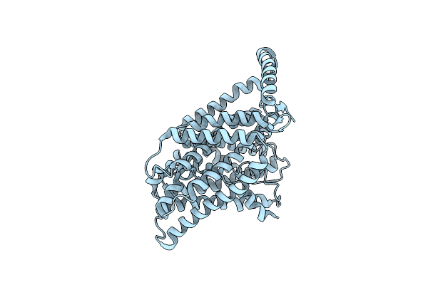 |
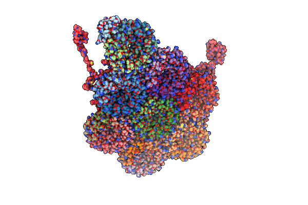
Organism: Homo sapiens
Method: ELECTRON MICROSCOPY Release Date: 2025-04-02 Classification: MEMBRANE PROTEIN |
 |
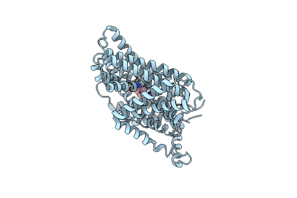
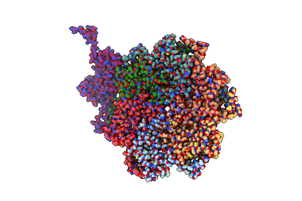
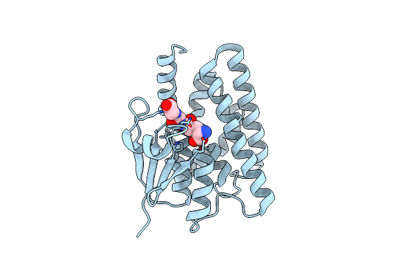
Organism: Homo sapiens
Method: ELECTRON MICROSCOPY Release Date: 2025-04-02 Classification: MEMBRANE PROTEIN Ligands: ARG |
 |
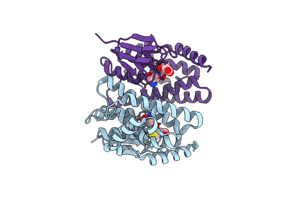
Organism: Homo sapiens
Method: ELECTRON MICROSCOPY Release Date: 2025-03-12 Classification: MEMBRANE PROTEIN Ligands: ARG |
 |
Organism: Homo sapiens
Method: ELECTRON MICROSCOPY Release Date: 2025-01-22 Classification: MEMBRANE PROTEIN |
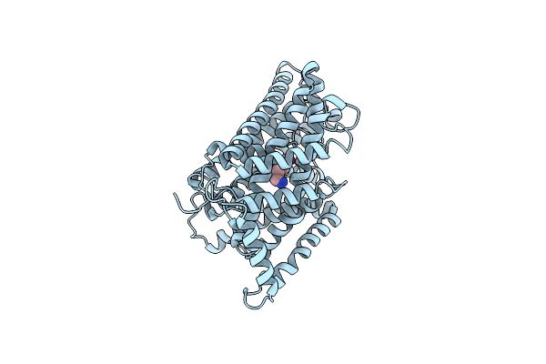 |
Organism: Homo sapiens
Method: ELECTRON MICROSCOPY Release Date: 2025-01-22 Classification: MEMBRANE PROTEIN Ligands: LYS |
 |
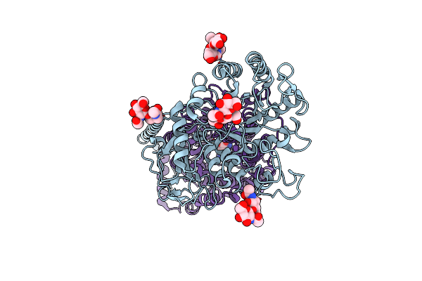
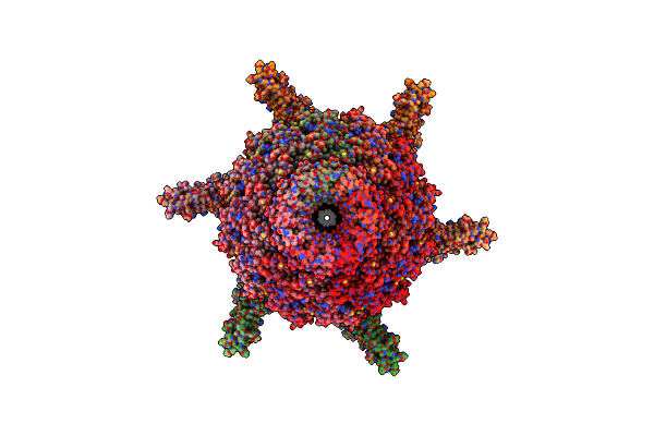
Cryo-Em Structure Of The Alpha-Carboxysome Shell Vertex From Prochlorococcus Med4
Organism: Prochlorococcus sp. med4
Method: ELECTRON MICROSCOPY Release Date: 2024-01-31 Classification: PHOTOSYNTHESIS |
 |
Organism: Prochlorococcus phage p-scsp1u
Method: ELECTRON MICROSCOPY Release Date: 2023-11-01 Classification: VIRUS |
 |
Organism: Prochlorococcus phage p-scsp1u
Method: ELECTRON MICROSCOPY Release Date: 2023-10-25 Classification: VIRUS |
 |
Organism: Synechococcus phage syn19
Method: X-RAY DIFFRACTION Resolution:1.70 Å Release Date: 2023-02-08 Classification: TRANSPORT PROTEIN Ligands: PO4, GOL |
 |
Organism: Prochlorococcus phage p-ssm2
Method: X-RAY DIFFRACTION Resolution:2.25 Å Release Date: 2023-02-08 Classification: TRANSPORT PROTEIN Ligands: PO4 |
 |
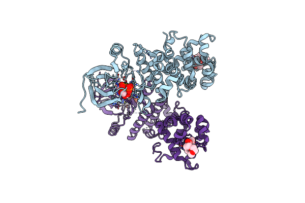
Organism: Homo sapiens
Method: ELECTRON MICROSCOPY Release Date: 2022-12-28 Classification: IMMUNE SYSTEM |
 |
Organism: Homo sapiens
Method: ELECTRON MICROSCOPY Release Date: 2022-12-28 Classification: IMMUNE SYSTEM Ligands: WJ6 |
 |
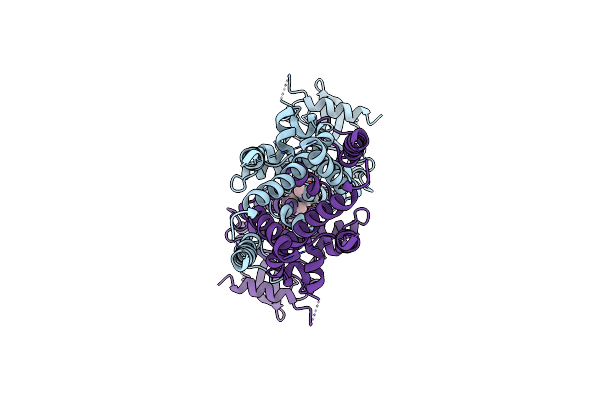
Crystal Structure Of A Glutathione S-Transferase Tau1 From Pinus Densata In Complex With Gsh
Organism: Pinus densata
Method: X-RAY DIFFRACTION Resolution:2.19 Å Release Date: 2022-12-07 Classification: TRANSFERASE Ligands: GSH |
 |
Organism: Streptococcus thermophilus lmg 18311
Method: X-RAY DIFFRACTION Resolution:2.96 Å Release Date: 2021-07-21 Classification: TRANSFERASE Ligands: SO4 |
 |
Organism: Microhyla heymonsi
Method: X-RAY DIFFRACTION Resolution:2.20 Å Release Date: 2021-04-21 Classification: ANTIMICROBIAL PROTEIN Ligands: CL |
 |
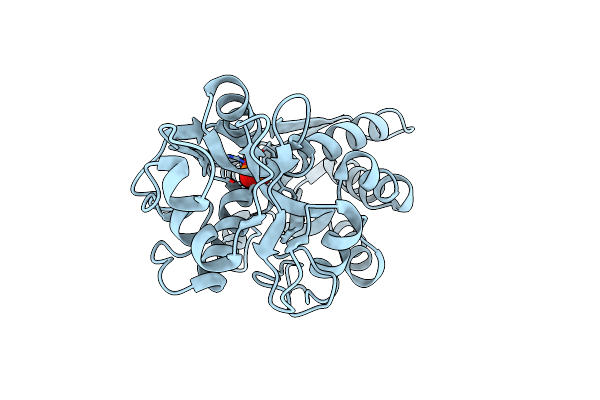
Crystal Structure Of A Glutathione S-Transferase Ptgstu30 From Populus Trichocarpa In Complex With Gsh
Organism: Populus trichocarpa
Method: X-RAY DIFFRACTION Resolution:1.25 Å Release Date: 2017-04-12 Classification: TRANSFERASE Ligands: GSH |
 |
Crystal Structure Of The R39W Mutant Of Populus Trichocarpa Glutathione Transferase Ptgstu30 In Complex With Glutathione
Organism: Populus trichocarpa
Method: X-RAY DIFFRACTION Resolution:2.63 Å Release Date: 2017-04-12 Classification: TRANSFERASE Ligands: GSH |
 |
Crystal Structure Of Mouse Hepatitis Virus Strain Dvim Hemagglutinin-Esterase
Organism: Murine coronavirus
Method: X-RAY DIFFRACTION Resolution:2.00 Å Release Date: 2016-05-11 Classification: VIRAL PROTEIN |
 |
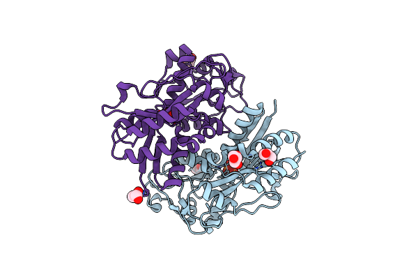
Crystal Structure Of Rat Coronavirus Strain New-Jersey Hemagglutinin-Esterase In Complex With 4N-Acetyl Sialic Acid
Organism: Rat coronavirus
Method: X-RAY DIFFRACTION Resolution:1.85 Å Release Date: 2016-05-11 Classification: VIRAL PROTEIN Ligands: NAG, NA, ZN, 6KL |
 |
Organism: Mus musculus
Method: X-RAY DIFFRACTION Resolution:2.90 Å Release Date: 2014-09-03 Classification: TRANSFERASE,HYDROLASE/INHIBITOR Ligands: 31J, ADP, MG |