
Search Count: 139
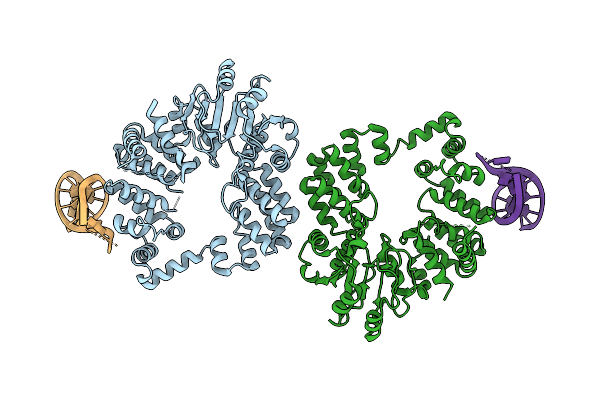 |
Organism: Thermus phage philo
Method: ELECTRON MICROSCOPY Release Date: 2025-12-10 Classification: RNA BINDING PROTEIN/RNA |
 |
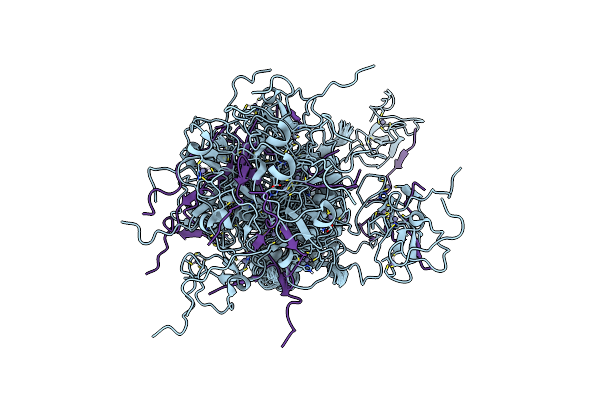
Organism: Thermus phage philo
Method: X-RAY DIFFRACTION Release Date: 2025-10-01 Classification: RNA BINDING PROTEIN Ligands: CA |
 |
Crystal Structure Of Dehydrogenase/Isomerase Fabx From Helicobacter Pylori In Complex With Inhibitor 1991
Organism: Helicobacter pylori
Method: X-RAY DIFFRACTION Release Date: 2025-08-20 Classification: BIOSYNTHETIC PROTEIN Ligands: A1ECY, FMN, SF4, MG |
 |
Crystal Structure Of Dehydrogenase/Isomerase Fabx From Helicobacter Pylori In Complex With Inhibitor 1872
Organism: Helicobacter pylori
Method: X-RAY DIFFRACTION Release Date: 2025-08-20 Classification: BIOSYNTHETIC PROTEIN Ligands: FMN, SF4, A1EEQ |
 |
Set Domain Of Histone-Lysine N-Methyltransferase Nsd2 In Complex With Selective Inhibitor
Organism: Homo sapiens
Method: SOLUTION NMR Release Date: 2025-04-30 Classification: TRANSFERASE/TRANSFERASE INHIBITOR Ligands: ZN, A1A0M |
 |
Organism: Homo sapiens
Method: X-RAY DIFFRACTION Resolution:1.92 Å Release Date: 2025-03-19 Classification: TRANSFERASE Ligands: ZN |
 |
Organism: Homo sapiens
Method: X-RAY DIFFRACTION Resolution:1.34 Å Release Date: 2025-03-19 Classification: TRANSFERASE Ligands: ZN |
 |
Organism: Homo sapiens
Method: X-RAY DIFFRACTION Resolution:2.40 Å Release Date: 2025-03-19 Classification: TRANSFERASE Ligands: ZN, SR |
 |
Nmr Solution Structures Of Ash1L Brd-Phd Domain In Complex With H3K4Me2 Peptide
Organism: Homo sapiens
Method: SOLUTION NMR Release Date: 2025-02-19 Classification: STRUCTURAL PROTEIN Ligands: ZN |
 |
Organism: Homo sapiens, Lama glama, Escherichia coli
Method: ELECTRON MICROSCOPY Release Date: 2024-07-24 Classification: MEMBRANE PROTEIN/IMMUNE SYSTEM Ligands: CLR |
 |
Organism: Homo sapiens, Lama glama, Escherichia coli
Method: ELECTRON MICROSCOPY Release Date: 2024-07-24 Classification: MEMBRANE PROTEIN/IMMUNE SYSTEM Ligands: 7LD |
 |
Organism: Homo sapiens, Lama glama, Escherichia coli
Method: ELECTRON MICROSCOPY Release Date: 2024-07-24 Classification: MEMBRANE PROTEIN/IMMUNE SYSTEM Ligands: A1D8X |
 |
Organism: Homo sapiens, Lama glama, Escherichia coli, Mus musculus
Method: ELECTRON MICROSCOPY Release Date: 2024-07-24 Classification: MEMBRANE PROTEIN/IMMUNE SYSTEM Ligands: A1D8X |
 |
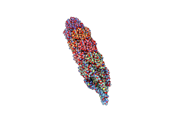
Organism: Caulobacter vibrioides
Method: ELECTRON MICROSCOPY Release Date: 2024-05-15 Classification: PROTEIN FIBRIL |
 |
Organism: Caulobacter phage phicb5, Caulobacter vibrioides
Method: ELECTRON MICROSCOPY Release Date: 2024-05-15 Classification: RECOMBINATION |
 |
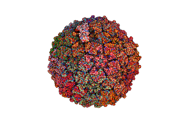
Organism: Caulobacter phage phicb5
Method: ELECTRON MICROSCOPY Release Date: 2024-05-15 Classification: VIRUS Ligands: CA |
 |
Organism: Pseudomonas aeruginosa pao1
Method: ELECTRON MICROSCOPY Release Date: 2024-03-13 Classification: MEMBRANE PROTEIN |
 |
Organism: Pseudomonas aeruginosa pao1, Pseudomonas phage pp7
Method: ELECTRON MICROSCOPY Release Date: 2024-03-13 Classification: MEMBRANE PROTEIN |
 |
Organism: Pseudomonas phage pp7
Method: ELECTRON MICROSCOPY Release Date: 2024-03-13 Classification: VIRAL PROTEIN |
 |
Organism: Acinetobacter genomosp. 16bj
Method: ELECTRON MICROSCOPY Release Date: 2024-03-06 Classification: CELL ADHESION |