
Search Count: 136
 |
Organism: Toxoplasma gondii
Method: ELECTRON MICROSCOPY Release Date: 2025-12-03 Classification: STRUCTURAL PROTEIN Ligands: GTP, MG, GDP |
 |
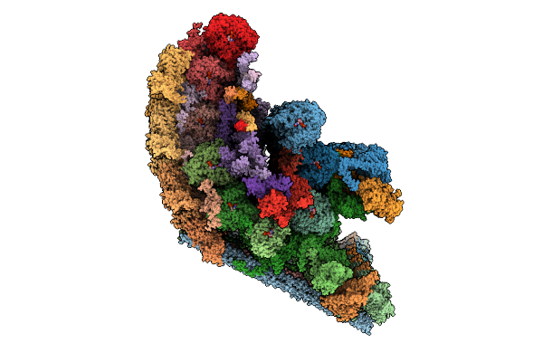
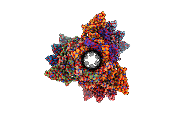
Cryo-Em Structure Of Intraconoidal Microtubule 2 (Icmt2) From Toxoplasma Gondii (8-Nm Repeat)
Organism: Toxoplasma gondii
Method: ELECTRON MICROSCOPY Release Date: 2025-12-03 Classification: STRUCTURAL PROTEIN Ligands: GTP, MG, GDP |
 |
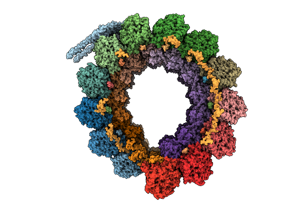
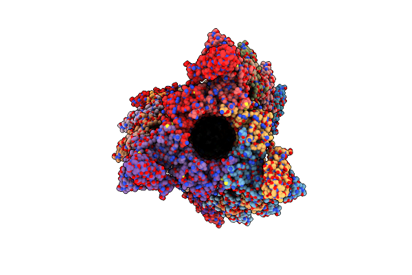
Cryo-Em Structure Of Intraconoidal Microtubule 1 (Icmt1) From Toxoplasma Gondii (8-Nm Repeat)
Organism: Toxoplasma gondii
Method: ELECTRON MICROSCOPY Release Date: 2025-12-03 Classification: STRUCTURAL PROTEIN Ligands: GTP, MG, GDP |
 |
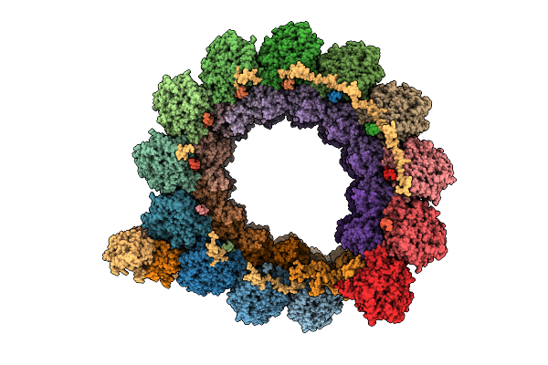
Cryo-Em Structure Of The Apical Region Of Subpellicular Microtubule (Spmt) From Toxoplasma Gondii (8-Nm Repeat)
Organism: Toxoplasma gondii
Method: ELECTRON MICROSCOPY Release Date: 2025-12-03 Classification: STRUCTURAL PROTEIN Ligands: GDP, GTP, MG |
 |
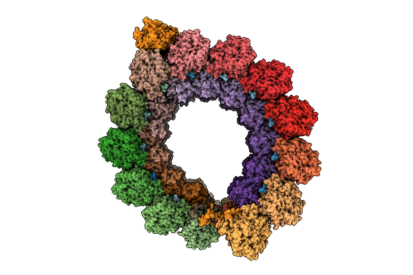
Organism: Toxoplasma gondii
Method: ELECTRON MICROSCOPY Release Date: 2025-11-19 Classification: STRUCTURAL PROTEIN |
 |
Organism: Bos taurus
Method: ELECTRON MICROSCOPY Release Date: 2025-03-26 Classification: STRUCTURAL PROTEIN Ligands: GTP, MG, GDP |
 |
Organism: Leishmania tarentolae
Method: ELECTRON MICROSCOPY Release Date: 2025-03-12 Classification: STRUCTURAL PROTEIN Ligands: GDP, MG, GTP, ZN, CA, ATP |
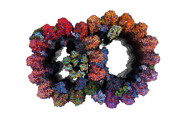 |
Atomic Model Of Bovine Fallopian Tube Cilia Doublet Microtubule (48-Nm Periodicity)
Organism: Bos taurus
Method: ELECTRON MICROSCOPY Release Date: 2024-10-30 Classification: STRUCTURAL PROTEIN Ligands: GDP, MG, GTP |
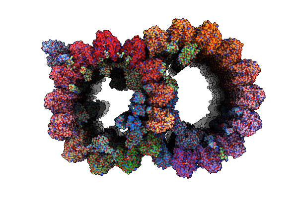 |
Atomic Model Of Porcine Brain Ventricles Cilia Doublet Microtubule (48-Nm Periodicity)
Organism: Sus scrofa
Method: ELECTRON MICROSCOPY Release Date: 2024-10-23 Classification: STRUCTURAL PROTEIN Ligands: GTP, GDP |
 |
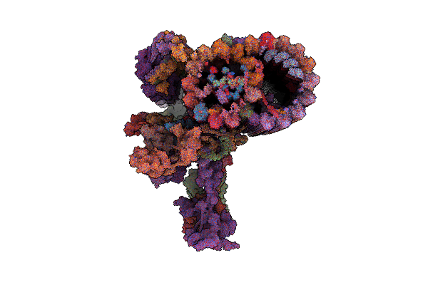
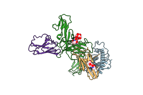
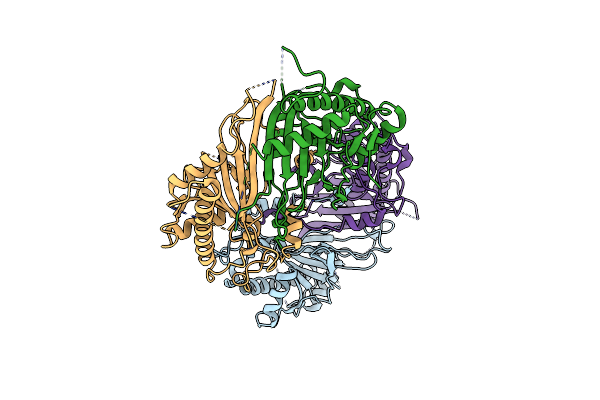
The Cryo-Em Structure Of Il-12, Receptor Subunit Beta-1 And Receptor Subunit Beta-2 Complex
Organism: Homo sapiens
Method: ELECTRON MICROSCOPY Release Date: 2024-07-24 Classification: CYTOKINE Ligands: NAG |
 |
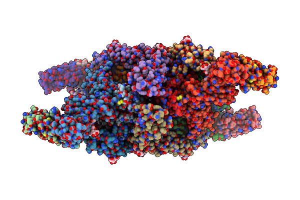
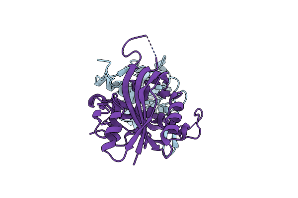
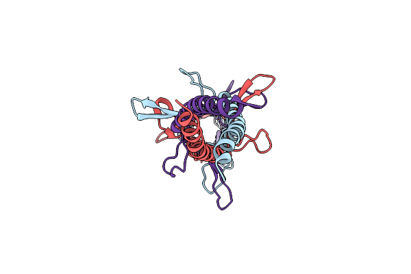
The Cryo-Em Structure Of Il-12, Receptor Subunit Beta-1 And Receptor Subunit Beta-2 Complex, Local Refinement
Organism: Homo sapiens
Method: ELECTRON MICROSCOPY Release Date: 2024-07-24 Classification: CYTOKINE Ligands: NAG |
 |
Organism: Vibrio phage icp1_2011_a
Method: X-RAY DIFFRACTION Resolution:2.56 Å Release Date: 2024-07-10 Classification: RNA BINDING PROTEIN |
 |
Organism: Vibrio phage icp1_2011_a
Method: X-RAY DIFFRACTION Resolution:2.37 Å Release Date: 2024-07-10 Classification: RNA BINDING PROTEIN |
 |
Organism: Vibrio phage icp1_2011_a
Method: X-RAY DIFFRACTION Resolution:3.00 Å Release Date: 2024-07-10 Classification: RNA BINDING PROTEIN/RNA |
 |
Organism: Oryza sativa
Method: X-RAY DIFFRACTION Resolution:3.16 Å Release Date: 2023-12-06 Classification: OXIDOREDUCTASE |
 |
Organism: Bos taurus
Method: ELECTRON MICROSCOPY Release Date: 2023-11-22 Classification: STRUCTURAL PROTEIN Ligands: GTP, MG, GDP |
 |
Organism: Escherichia phage lambda
Method: ELECTRON MICROSCOPY Release Date: 2023-10-18 Classification: VIRAL PROTEIN |
 |
Organism: Escherichia phage lambda
Method: ELECTRON MICROSCOPY Release Date: 2023-10-18 Classification: VIRAL PROTEIN Ligands: SF4 |
 |
Organism: Escherichia phage lambda
Method: ELECTRON MICROSCOPY Release Date: 2023-10-18 Classification: VIRAL PROTEIN Ligands: SF4 |
 |
Organism: Escherichia phage lambda
Method: ELECTRON MICROSCOPY Release Date: 2023-10-18 Classification: VIRAL PROTEIN |