
Search Count: 18
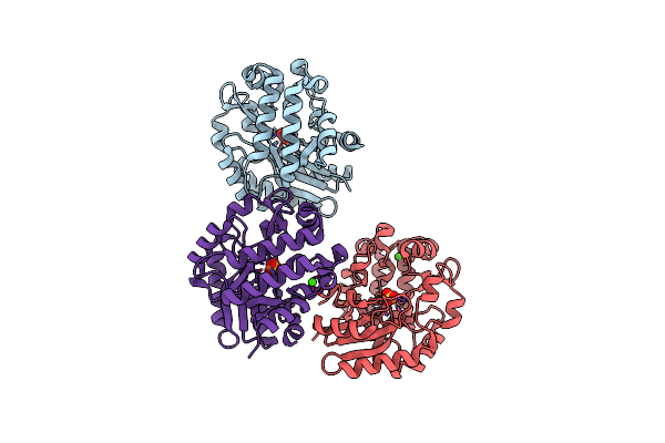 |
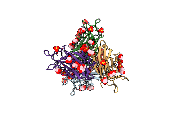
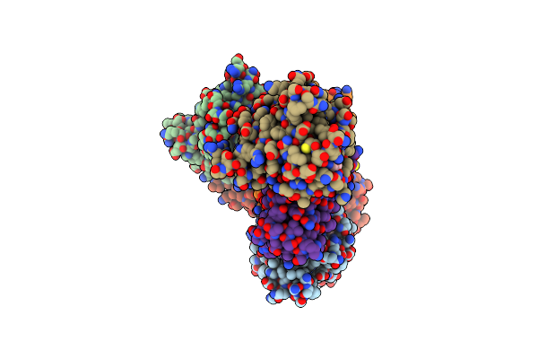
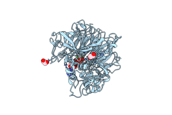
Crystal Structure Of The Putative Histidinol Phosphatase Hisk From Listeria Monocytogenes With Trinuclear Metals Determined By Pixe Revealing Sulphate Ion In Active Site. Based On Pixe Analysis And Original Date From 3Dcp
Organism: Listeria monocytogenes serotype 4b str. h7858
Method: X-RAY DIFFRACTION Resolution:2.10 Å Release Date: 2019-12-25 Classification: HYDROLASE Ligands: MN, CO, SO4, FE, CA |
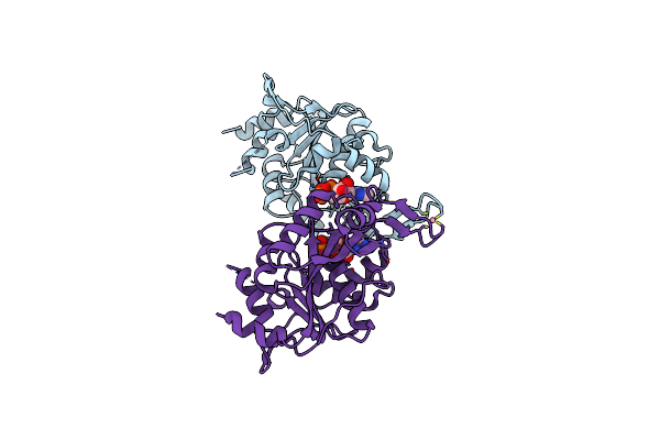 |
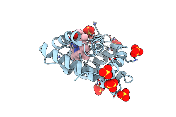
The Nucleotide-Binding Protein Af_226 In Complex With Adp From Archaeoglobus Fulgidus With Co Found By Pixe. Based On 3Kb1.
Organism: Archaeoglobus fulgidus
Method: X-RAY DIFFRACTION Resolution:2.87 Å Release Date: 2019-12-25 Classification: METAL TRANSPORT Ligands: ADP, CO |
 |
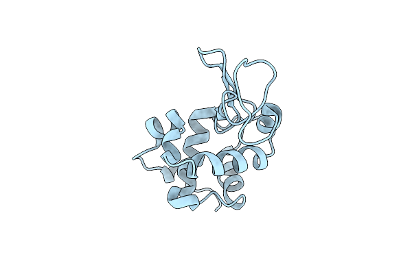
X-Ray Structure Of The C-Terminal Domain (277-440) Of Putative Chitobiase From Bacteroides Thetaiotaomicron. Northeast Structural Genomics Consortium Target Btr324A. Re-Refinement Of 3Ggl With Correct Metal Mn Replacing Zn. New Metal Confirmed With Pixe Analysis Of Original Sample.
Organism: Bacteroides thetaiotaomicron
Method: X-RAY DIFFRACTION Resolution:3.00 Å Release Date: 2019-12-25 Classification: HYDROLASE Ligands: PEG, MN |
 |
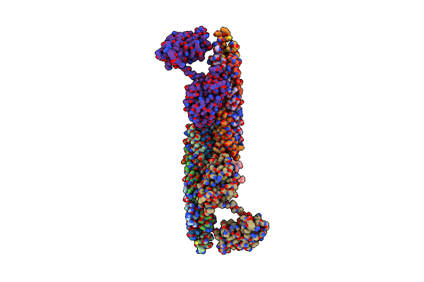
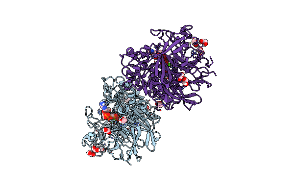
Structure Of The Ca2+-Bound Synaptotagmin-1 Snare Complex (Long Unit Cell Form) - From Xfel Diffraction
Organism: Rattus norvegicus
Method: X-RAY DIFFRACTION Resolution:3.50 Å Release Date: 2016-10-19 Classification: ENDOCYTOSIS,EXOCYTOSIS Ligands: CA |
 |
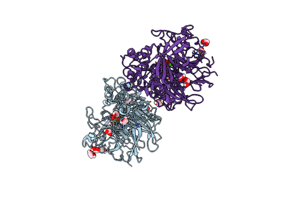
Structure Of The Ca2+-Bound Synaptotagmin-1 Snare Complex (Long Unit Cell Form) - From Synchrotron Diffraction
Organism: Rattus norvegicus
Method: X-RAY DIFFRACTION Resolution:4.10 Å Release Date: 2016-10-19 Classification: ENDOCYTOSIS,EXOCYTOSIS Ligands: CA |
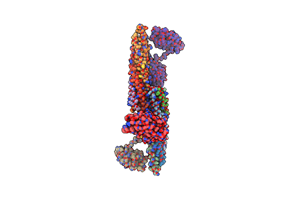 |
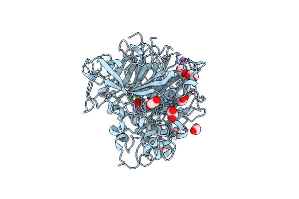
Structure Of The Ca2+-Bound Synaptotagmin-1 Snare Complex (Long Unit Cell Form)
Organism: Rattus norvegicus
Method: X-RAY DIFFRACTION Resolution:3.50 Å Release Date: 2015-08-12 Classification: ENDOCYTOSIS,EXOCYTOSIS Ligands: CA |
 |
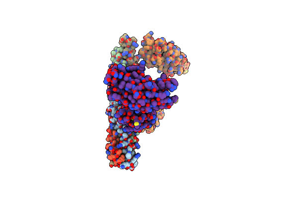
Structure Of The Ca2+-Bound Synaptotagmin-1 Snare Complex (Short Unit Cell Form)
Organism: Rattus norvegicus
Method: X-RAY DIFFRACTION Resolution:3.60 Å Release Date: 2015-08-12 Classification: ENDOCYTOSIS,EXOCYTOSIS Ligands: CA |
 |
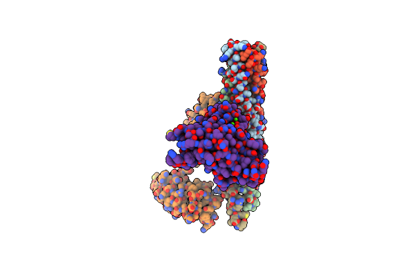
Structure Of The Mg2+-Bound Synaptotagmin-1 Snare Complex (Short Unit Cell Form)
Organism: Rattus norvegicus
Method: X-RAY DIFFRACTION Resolution:4.10 Å Release Date: 2015-08-12 Classification: ENDOCYTOSIS,EXOCYTOSIS Ligands: MG |
 |
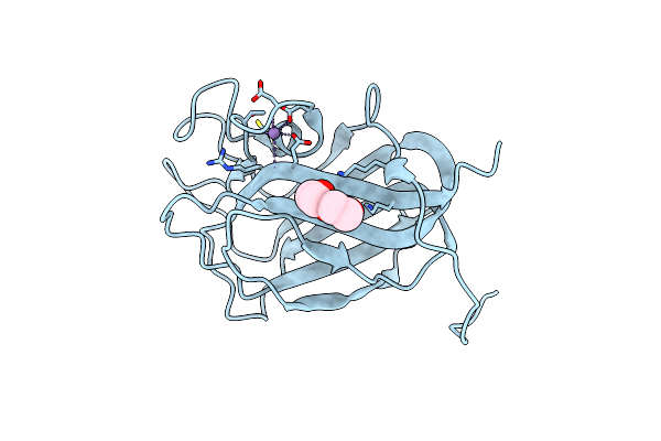
Crystal Structure Of The Quintuple Mutant Of The Synaptotagmin-1 C2B Domain
Organism: Rattus norvegicus
Method: X-RAY DIFFRACTION Resolution:1.65 Å Release Date: 2015-08-12 Classification: SIGNALING PROTEIN Ligands: GOL, SO4 |
 |
Structure Of Hen Egg-White Lysozyme From A Microfludic Harvesting Device Using Synchrotron Radiation (2.5A)
Organism: Gallus gallus
Method: X-RAY DIFFRACTION Resolution:2.50 Å Release Date: 2015-04-22 Classification: HYDROLASE |
 |
Recombinant Sperm Whale P6 Myoglobin Solved With Single Pulse Free Electron Laser Data
Organism: Physeter catodon
Method: X-RAY DIFFRACTION Resolution:1.36 Å Release Date: 2014-11-05 Classification: OXYGEN TRANSPORT Ligands: HEM, SO4 |
 |
Organism: Homo sapiens
Method: X-RAY DIFFRACTION Resolution:2.80 Å Release Date: 2014-06-04 Classification: TRANSPORT PROTEIN Ligands: GCP, MG |
 |
Organism: Homo sapiens
Method: X-RAY DIFFRACTION Resolution:2.97 Å Release Date: 2014-06-04 Classification: SIGNALING PROTEIN Ligands: GCP, MG |
 |
Organism: Pseudomonas fluorescens
Method: X-RAY DIFFRACTION Resolution:1.52 Å Release Date: 2013-03-20 Classification: HYDROLASE Ligands: ACP, CL, LI, CA, FEO, EDO |
 |
Organism: Pseudomonas fluorescens
Method: X-RAY DIFFRACTION Resolution:1.25 Å Release Date: 2013-03-13 Classification: HYDROLASE Ligands: PO4, CA, FEO, EDO, CL |
 |
Organism: Pseudomonas fluorescens
Method: X-RAY DIFFRACTION Resolution:1.10 Å Release Date: 2012-12-05 Classification: HYDROLASE Ligands: CL, LI, NA, EDO, CA, FEO |
 |
Organism: Pseudomonas fluorescens
Method: X-RAY DIFFRACTION Resolution:1.79 Å Release Date: 2012-12-05 Classification: HYDROLASE Ligands: EDO, ACP, CA, FEO |
 |
Pseudomonas Fluorescens Phox In Complex With Vanadate, A Transition State Analogue
Organism: Pseudomonas fluorescens
Method: X-RAY DIFFRACTION Resolution:1.39 Å Release Date: 2012-08-08 Classification: HYDROLASE Ligands: VO4, CL, CA, FEO |