
Search Count: 25
 |
Organism: Homo sapiens
Method: ELECTRON MICROSCOPY Release Date: 2024-11-27 Classification: STRUCTURAL PROTEIN Ligands: GDP, GTP, MG |
 |
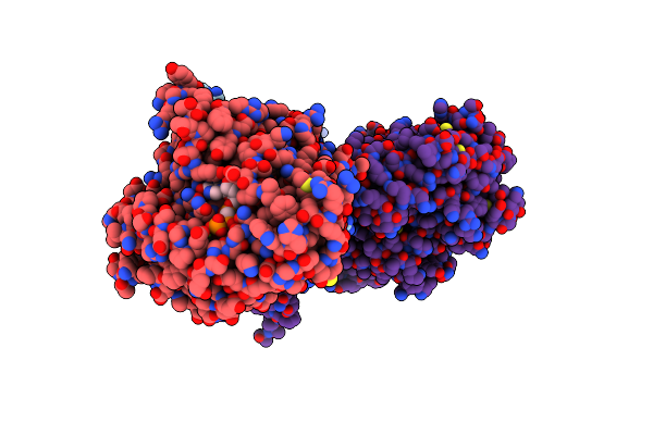
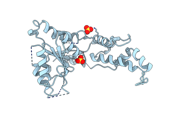
Organism: Homo sapiens
Method: X-RAY DIFFRACTION Resolution:1.80 Å Release Date: 2024-07-17 Classification: HYDROLASE Ligands: MLT, IMD, ZN |
 |
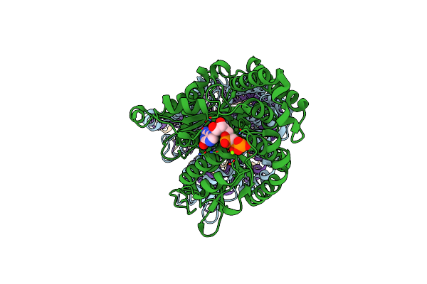
Organism: Homo sapiens
Method: X-RAY DIFFRACTION Resolution:2.30 Å Release Date: 2024-07-17 Classification: HYDROLASE/INHIBITOR Ligands: ZN, MLT, A1AAG |
 |
Organism: Homo sapiens
Method: X-RAY DIFFRACTION Resolution:2.30 Å Release Date: 2024-07-17 Classification: HYDROLASE/INHIBITOR Ligands: ZN, MLT, K |
 |
Organism: Homo sapiens
Method: X-RAY DIFFRACTION Resolution:2.36 Å Release Date: 2024-07-17 Classification: HYDROLASE Ligands: ZN |
 |
Organism: Homo sapiens, Sus scrofa
Method: ELECTRON MICROSCOPY Release Date: 2024-07-17 Classification: HYDROLASE/SUBSTRATE Ligands: ZN, GLU |
 |
Organism: Homo sapiens, Sus scrofa
Method: ELECTRON MICROSCOPY Release Date: 2024-07-17 Classification: HYDROLASE/SUBSTRATE Ligands: ZN, GLU |
 |
Organism: Homo sapiens, Sus scrofa
Method: ELECTRON MICROSCOPY Release Date: 2024-07-17 Classification: HYDROLASE/SUBSTRATE Ligands: ZN, GLU |
 |
Organism: Homo sapiens, Sus scrofa
Method: ELECTRON MICROSCOPY Release Date: 2024-07-17 Classification: HYDROLASE/SUBSTRATE,STRUCTURAL PROTEIN Ligands: MG, GTP, G2P, ZN, GLU |
 |
Organism: Homo sapiens, Sus scrofa
Method: ELECTRON MICROSCOPY Release Date: 2024-07-17 Classification: HYDROLASE/SUBSTRATE,STRUCTURAL PROTEIN Ligands: MG, GTP, G2P, GLU, ZN |
 |
Organism: Homo sapiens, Sus scrofa
Method: ELECTRON MICROSCOPY Release Date: 2024-07-17 Classification: HYDROLASE/SUBSTRATE,STRUCTURAL PROTEIN Ligands: MG, GTP, G2P, GLU, ZN |
 |
Organism: Homo sapiens
Method: ELECTRON MICROSCOPY Release Date: 2024-06-12 Classification: STRUCTURAL PROTEIN Ligands: GTP, MG, G2P |
 |
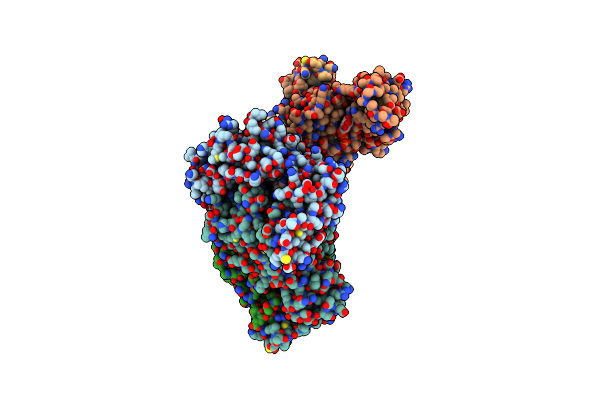
Organism: Mus musculus, Homo sapiens
Method: ELECTRON MICROSCOPY Release Date: 2024-05-29 Classification: LIGASE Ligands: GTP, MG, G2P, ATP |
 |
Organism: Mus musculus, Homo sapiens
Method: ELECTRON MICROSCOPY Release Date: 2024-05-08 Classification: LIGASE Ligands: GTP, MG, G2P |
 |
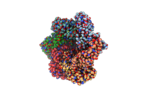
Organism: Caenorhabditis elegans, Synthetic construct
Method: ELECTRON MICROSCOPY Release Date: 2019-10-09 Classification: MOTOR PROTEIN Ligands: ATP, MG |
 |
Organism: Caenorhabditis elegans, Synthetic construct
Method: ELECTRON MICROSCOPY Release Date: 2019-10-09 Classification: MOTOR PROTEIN Ligands: ATP, MG |
 |
Katanin Hexamer In The Ring Conformation With Resolved Protomer One In Complex With Substrate
Organism: Caenorhabditis elegans, Synthetic construct
Method: ELECTRON MICROSCOPY Release Date: 2019-10-09 Classification: MOTOR PROTEIN Ligands: ATP, MG |
 |
Organism: Drosophila melanogaster
Method: ELECTRON MICROSCOPY Release Date: 2019-06-12 Classification: MOTOR PROTEIN Ligands: ATP, MG, ADP |
 |
Organism: Caenorhabditis elegans
Method: ELECTRON MICROSCOPY Release Date: 2017-08-09 Classification: MOTOR PROTEIN Ligands: ATP |
 |
Organism: Caenorhabditis elegans
Method: X-RAY DIFFRACTION Resolution:3.30 Å Release Date: 2017-08-09 Classification: CYTOSOLIC PROTEIN Ligands: SO4 |