
Search Count: 63
 |
Organism: Nonlabens marinus s1-08
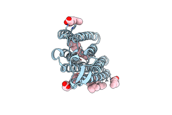
Method: X-RAY DIFFRACTION Resolution:1.85 Å Release Date: 2021-04-14 Classification: MEMBRANE PROTEIN Ligands: CL, RET, OLA |
 |
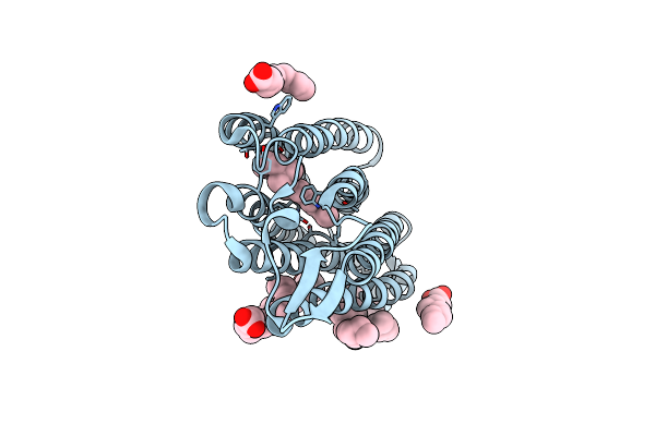
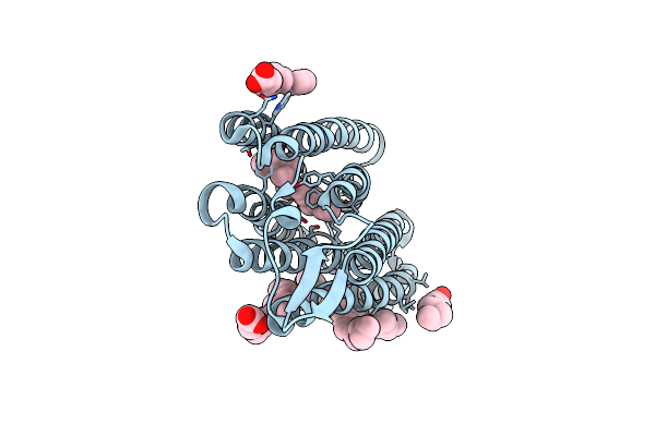
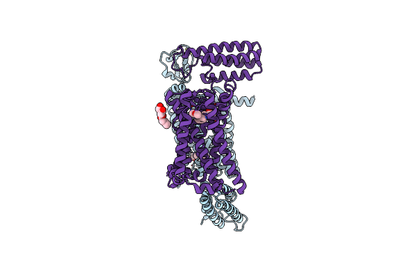
Structure Of Chloride Ion Pumping Rhodopsin (Clr) With Ntq Motif 50 Ps After Light Activation
Organism: Nonlabens marinus s1-08
Method: X-RAY DIFFRACTION Resolution:1.85 Å Release Date: 2021-04-14 Classification: MEMBRANE PROTEIN Ligands: CL, RET, OLA |
 |
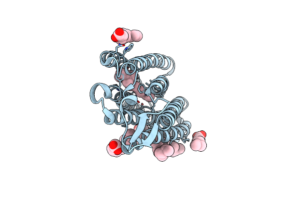
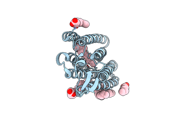
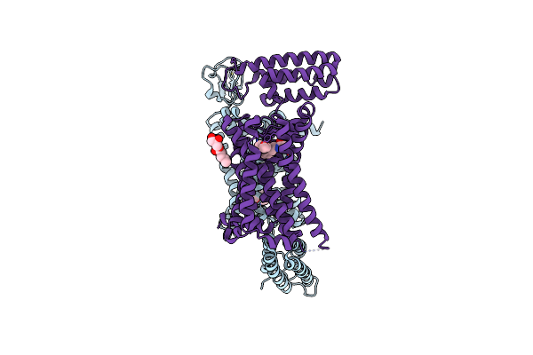
Structure Of Chloride Ion Pumping Rhodopsin (Clr) With Ntq Motif 100 Ps After Light Activation (0.90Mj/Mm2)
Organism: Nonlabens marinus s1-08
Method: X-RAY DIFFRACTION Resolution:1.85 Å Release Date: 2021-04-14 Classification: MEMBRANE PROTEIN Ligands: CL, RET, OLA |
 |
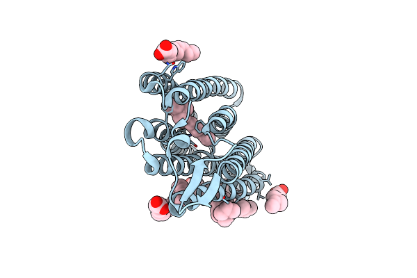
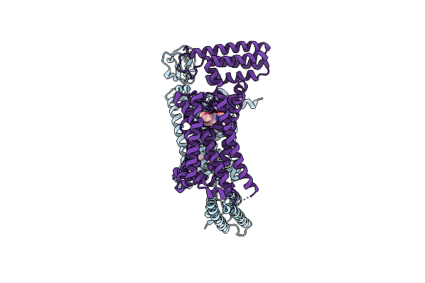
Structure Of Chloride Ion Pumping Rhodopsin (Clr) With Ntq Motif 100 Ps After Light Activation (0.17Mj/Mm2)
Organism: Nonlabens marinus s1-08
Method: X-RAY DIFFRACTION Resolution:1.85 Å Release Date: 2021-04-14 Classification: MEMBRANE PROTEIN Ligands: CL, RET, OLA |
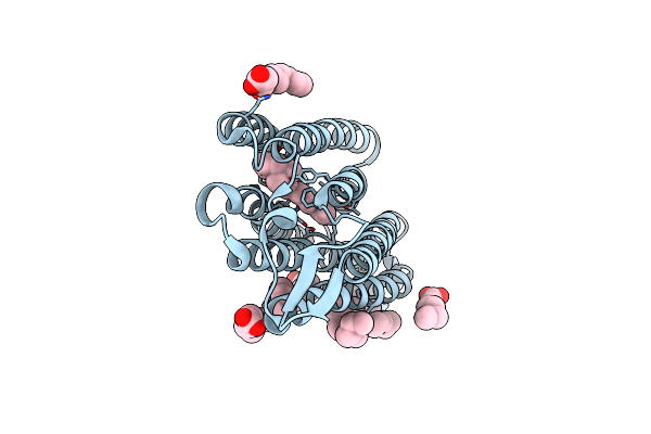 |
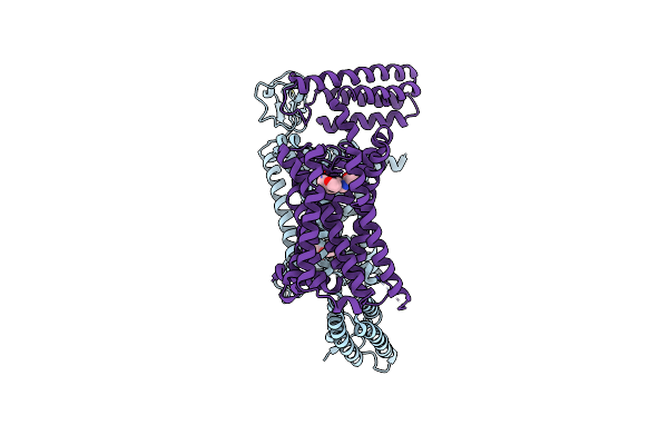
Structure Of Chloride Ion Pumping Rhodopsin (Clr) With Ntq Motif 100 Ps After Light Activation (2.63Mj/Mm2)
Organism: Nonlabens marinus s1-08
Method: X-RAY DIFFRACTION Resolution:1.85 Å Release Date: 2021-04-14 Classification: MEMBRANE PROTEIN Ligands: CL, RET, OLA |
 |
Structure Of Chloride Ion Pumping Rhodopsin (Clr) With Ntq Motif 100 Ps After Light Activation (6.49 Mj/Mm2)
Organism: Nonlabens marinus s1-08
Method: X-RAY DIFFRACTION Resolution:1.85 Å Release Date: 2021-04-14 Classification: MEMBRANE PROTEIN Ligands: CL, RET, OLA |
 |
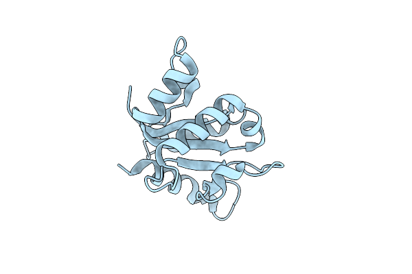
Organism: Homo sapiens
Method: ELECTRON CRYSTALLOGRAPHY Resolution:3.00 Å Release Date: 2021-03-10 Classification: PROTEIN BINDING |
 |
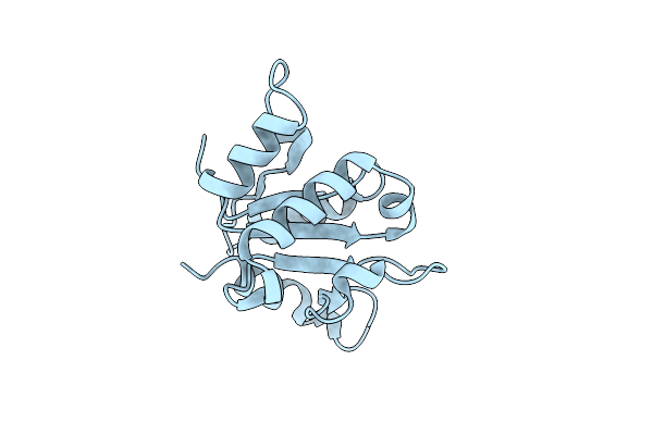
Sfx Structure Of The Myd88 Tir Domain Higher-Order Assembly (Solved, Rebuilt And Refined Using An Identical Protocol To The Microed Structure Of The Myd88 Tir Domain Higher-Order Assembly)
Organism: Homo sapiens
Method: X-RAY DIFFRACTION Resolution:2.30 Å Release Date: 2021-03-10 Classification: PROTEIN BINDING |
 |
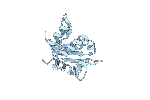
Organism: Homo sapiens
Method: X-RAY DIFFRACTION Resolution:2.30 Å Release Date: 2021-03-10 Classification: PROTEIN BINDING |
 |
Transitional Unit Cell 1 Of Adenine Riboswitch Aptamer Crystal Phase Transition Upon Ligand Binding
Organism: Vibrio vulnificus
Method: X-RAY DIFFRACTION Resolution:3.03 Å Release Date: 2020-11-11 Classification: RNA Ligands: ADE, MG |
 |
Transitional Unit Cell 2 Of Adenine Riboswitch Aptamer Crystal Phase Transition Upon Ligand Binding
Organism: Vibrio vulnificus
Method: X-RAY DIFFRACTION Resolution:3.00 Å Release Date: 2020-11-11 Classification: RNA Ligands: ADE, K, MG |
 |
Organism: Nonlabens marinus s1-08
Method: X-RAY DIFFRACTION Resolution:1.85 Å Release Date: 2020-09-30 Classification: MEMBRANE PROTEIN Ligands: CL, RET, OLA |
 |
Dark State Structure Of Chloride Ion Pumping Rhodopsin (Clr) With Ntq Motif
Organism: Nonlabens marinus s1-08
Method: X-RAY DIFFRACTION Resolution:1.65 Å Release Date: 2020-09-16 Classification: MEMBRANE PROTEIN Ligands: CL, RET, OLA |
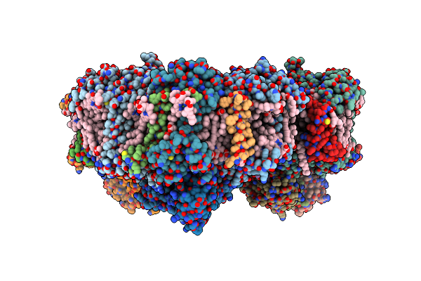 |
Membrane Protein Megahertz Crystallography At The European Xfel, Photosystem I Xfel At 2.9 A
Organism: Thermosynechococcus elongatus (strain bp-1)
Method: X-RAY DIFFRACTION Resolution:2.90 Å Release Date: 2019-11-27 Classification: PHOTOSYNTHESIS Ligands: CL0, CLA, PQN, SF4, BCR, LHG, LMG, CA |
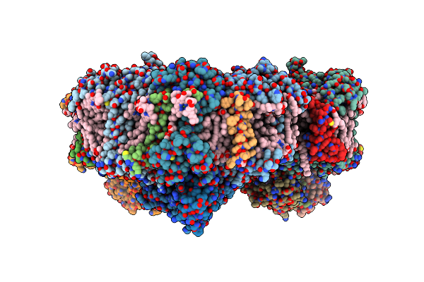 |
Membrane Protein Megahertz Crystallography At The European Xfel, Photosystem I At Synchrotron To 2.9 A
Organism: Thermosynechococcus elongatus (strain bp-1)
Method: X-RAY DIFFRACTION Resolution:2.90 Å Release Date: 2019-11-20 Classification: PHOTOSYNTHESIS Ligands: CL0, CLA, PQN, SF4, BCR, LHG, LMG, CA |
 |
Xfel Crystal Structure Of Human Melatonin Receptor Mt2 In Complex With 2-Phenylmelatonin
Organism: Escherichia coli, Homo sapiens, Clostridium pasteurianum
Method: X-RAY DIFFRACTION Resolution:2.80 Å Release Date: 2019-04-24 Classification: MEMBRANE PROTEIN Ligands: JEY, ZN, OLC |
 |
Xfel Crystal Structure Of Human Melatonin Receptor Mt2 (H208A) In Complex With 2-Phenylmelatonin
Organism: Escherichia coli, Homo sapiens, Clostridium pasteurianum
Method: X-RAY DIFFRACTION Resolution:3.20 Å Release Date: 2019-04-24 Classification: MEMBRANE PROTEIN Ligands: JEY, ZN, OLC |
 |
Xfel Crystal Structure Of Human Melatonin Receptor Mt2 (N86D) In Complex With 2-Phenylmelatonin
Organism: Escherichia coli, Homo sapiens, Clostridium pasteurianum
Method: X-RAY DIFFRACTION Resolution:3.10 Å Release Date: 2019-04-24 Classification: MEMBRANE PROTEIN Ligands: JEY, ZN |
 |
Xfel Crystal Structure Of Human Melatonin Receptor Mt2 In Complex With Ramelteon
Organism: Escherichia coli, Homo sapiens, Clostridium pasteurianum
Method: X-RAY DIFFRACTION Resolution:3.30 Å Release Date: 2019-04-24 Classification: MEMBRANE PROTEIN Ligands: JEV, ZN |
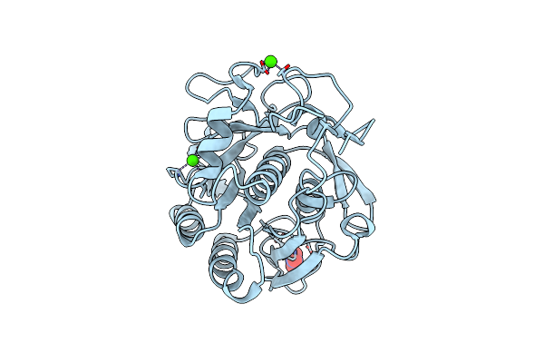 |
High-Viscosity Injector-Based Pink Beam Serial Crystallography Of Micro-Crystals At A Synchrotron Radiation Source.
Organism: Parengyodontium album
Method: X-RAY DIFFRACTION Resolution:1.80 Å Release Date: 2019-04-24 Classification: HYDROLASE Ligands: CA, NO3 |