
Search Count: 5
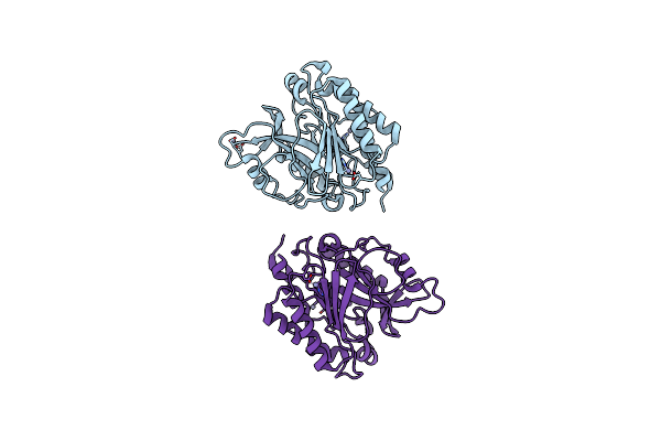 |
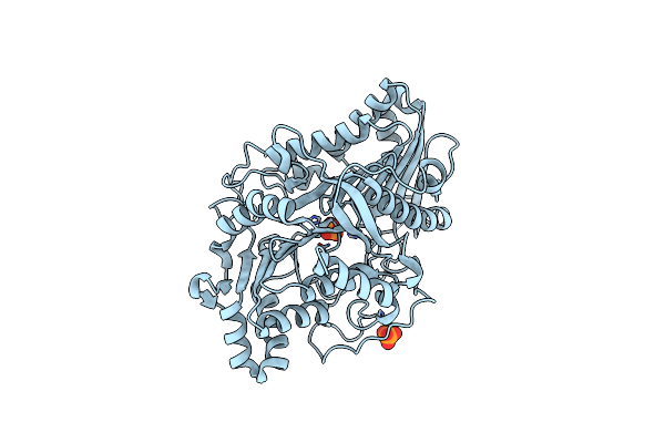
Crystal Structure Of The Y247S/Y251S Mutant Of Phosphatidylinositol-Specific Phospholipase C From Bacillus Thuringiensis
Organism: Bacillus thuringiensis
Method: X-RAY DIFFRACTION Resolution:1.75 Å Release Date: 2009-04-14 Classification: LYASE Ligands: ZN |
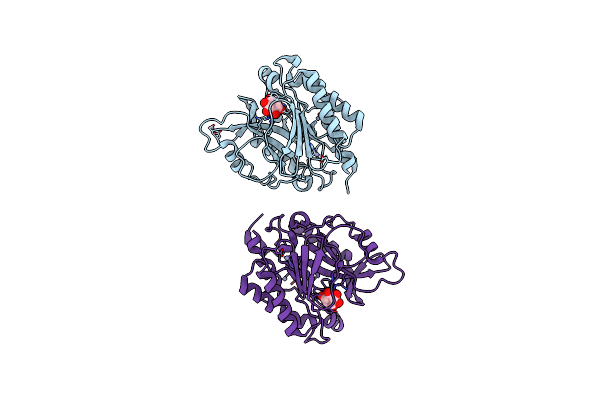 |
Crystal Structure Of The Myo-Inositol Bound Y247S/Y251S Mutant Of Phosphatidylinositol-Specific Phospholipase C From Bacillus Thuringiensis
Organism: Bacillus thuringiensis
Method: X-RAY DIFFRACTION Resolution:1.95 Å Release Date: 2009-04-14 Classification: LYASE Ligands: INS, ZN |
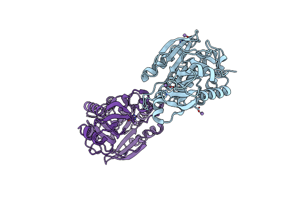 |
Crystal Structure Of The Y246S/Y247S/Y248S/Y251S Mutant Of Phosphatidylinositol-Specific Phospholipase C From Bacillus Thuringiensis
Organism: Bacillus thuringiensis
Method: X-RAY DIFFRACTION Resolution:1.78 Å Release Date: 2009-04-14 Classification: LYASE Ligands: MN |
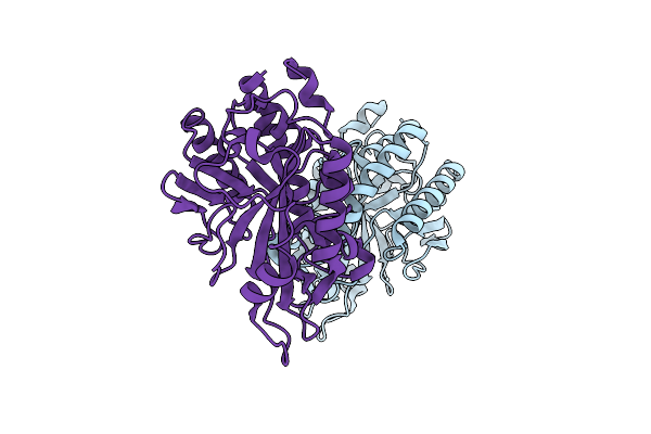 |
Structure Of The W47A/W242A Mutant Of Bacterial Phosphatidylinositol-Specific Phospholipase C
Organism: Bacillus thuringiensis
Method: X-RAY DIFFRACTION Resolution:1.84 Å Release Date: 2007-02-13 Classification: LYASE |
 |
Organism: Streptomyces sp.
Method: X-RAY DIFFRACTION Resolution:1.40 Å Release Date: 2001-05-16 Classification: HYDROLASE Ligands: PO4 |