
Search Count: 17
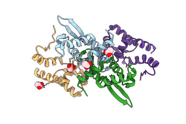 |
1.93 A Resolution X-Ray Crystal Structure Of The Transcriptional Regulator Srnr From Streptomyces Griseus
Organism: Streptomyces griseus
Method: X-RAY DIFFRACTION Resolution:1.93 Å Release Date: 2022-05-25 Classification: DNA BINDING PROTEIN Ligands: ACT, NA |
 |
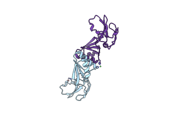
X-Ray Structure Of A Truncated Mutant Of The Metallochaperone Cooj With A High-Affinity Nickel-Binding Site
Organism: Rhodospirillum rubrum
Method: X-RAY DIFFRACTION Resolution:2.04 Å Release Date: 2019-03-27 Classification: METAL BINDING PROTEIN Ligands: NI, PEG, CL, CA, PGE, Z3P |
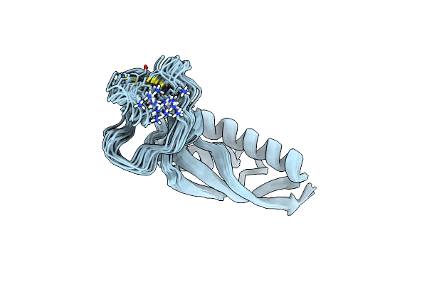 |
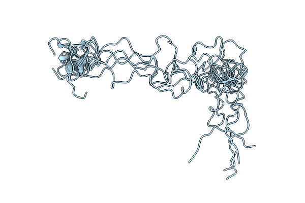
Solution Structure Of The Ni Metallochaperone Hypa From Helicobacter Pylori
Organism: Helicobacter pylori j99
Method: SOLUTION NMR Release Date: 2018-10-10 Classification: METAL BINDING PROTEIN Ligands: ZN |
 |
Crystal Structure Of The Apo-Form Of The Co Dehydrogenase Accessory Protein Coot From Rhodospirillum Rubrum
Organism: Rhodospirillum rubrum
Method: X-RAY DIFFRACTION Resolution:1.90 Å Release Date: 2017-05-10 Classification: nickel-binding protein |
 |
Organism: Trypanosoma brucei
Method: SOLUTION NMR Release Date: 2016-01-27 Classification: PROTEIN BINDING |
 |
Organism: Canavalia ensiformis
Method: SOLUTION NMR Release Date: 2015-01-28 Classification: HYDROLASE |
 |
Organism: Brucella suis
Method: X-RAY DIFFRACTION Resolution:1.85 Å Release Date: 2014-10-01 Classification: TRANSPORT PROTEIN Ligands: GOL, SO4 |
 |
Organism: Brucella suis
Method: X-RAY DIFFRACTION Resolution:1.95 Å Release Date: 2014-10-01 Classification: TRANSPORT PROTEIN Ligands: EDT, FE, SO4, GOL |
 |
Organism: Campylobacter jejuni
Method: X-RAY DIFFRACTION Resolution:2.40 Å Release Date: 2014-10-01 Classification: TRANSPORT PROTEIN Ligands: GOL |
 |
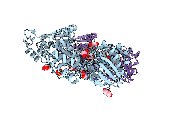
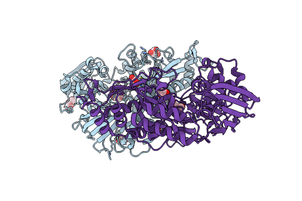
Crystal Structure Of Nikz From Campylobacter Jejuni In Complex With Ni(L-His)
Organism: Campylobacter jejuni
Method: X-RAY DIFFRACTION Resolution:2.20 Å Release Date: 2014-10-01 Classification: TRANSPORT PROTEIN Ligands: NI, HIS, MG, PEG, GOL |
 |
Crystal Structure Of Nikz From Campylobacter Jejuni In Complex With Ni(Ii) Ion
Organism: Campylobacter jejuni
Method: X-RAY DIFFRACTION Resolution:1.90 Å Release Date: 2014-10-01 Classification: TRANSPORT PROTEIN Ligands: NI, OXL, GOL |
 |
Organism: Yersinia pestis
Method: X-RAY DIFFRACTION Resolution:2.70 Å Release Date: 2014-10-01 Classification: TRANSPORT PROTEIN Ligands: NI, HIS |
 |
Organism: Yersinia pestis
Method: X-RAY DIFFRACTION Resolution:3.00 Å Release Date: 2014-10-01 Classification: TRANSPORT PROTEIN Ligands: NI |
 |
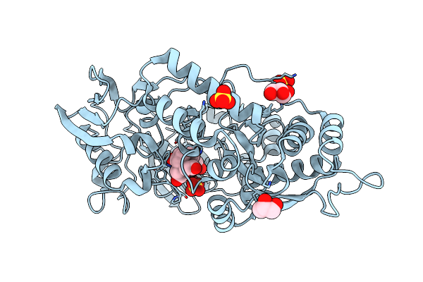
Organism: Sporosarcina pasteurii
Method: X-RAY DIFFRACTION Resolution:1.88 Å Release Date: 2013-10-09 Classification: CHAPERONE Ligands: NI, ZN |
 |
Organism: Helicobacter pylori
Method: X-RAY DIFFRACTION Resolution:1.59 Å Release Date: 2011-11-02 Classification: METAL BINDING PROTEIN Ligands: FMT, NI |
 |
Organism: Helicobacter pylori
Method: X-RAY DIFFRACTION Resolution:2.52 Å Release Date: 2011-11-02 Classification: METAL BINDING PROTEIN Ligands: ZN, FMT |
 |
Organism: Helicobacter pylori
Method: X-RAY DIFFRACTION Resolution:2.00 Å Release Date: 2011-11-02 Classification: METAL BINDING PROTEIN Ligands: SO4, CL |