
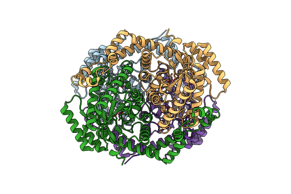
Search Count: 197
 |
Organism: Homo sapiens
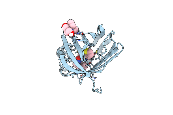
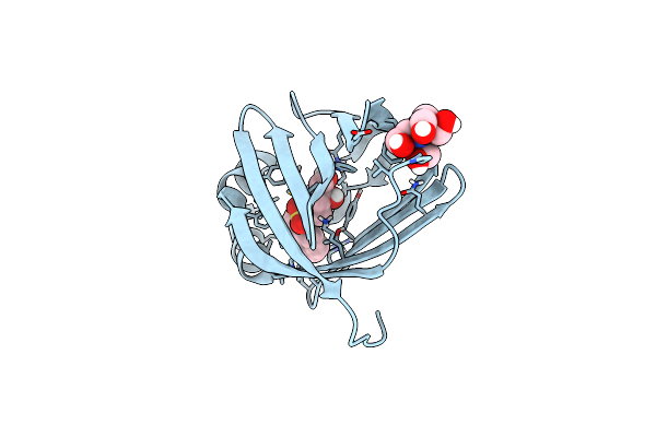
Method: X-RAY DIFFRACTION Release Date: 2025-10-22 Classification: ONCOPROTEIN Ligands: ZN, A1IWG |
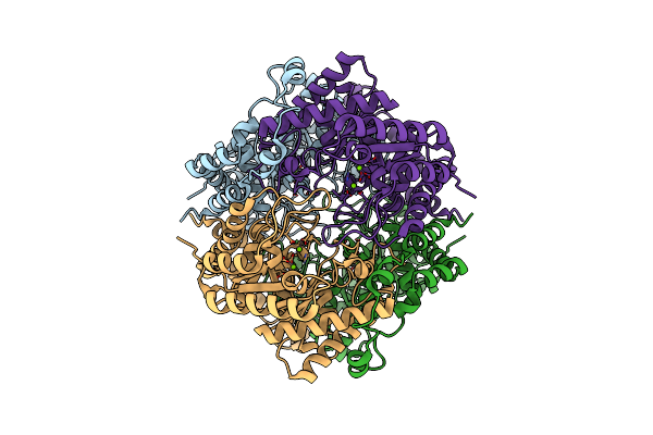
 |
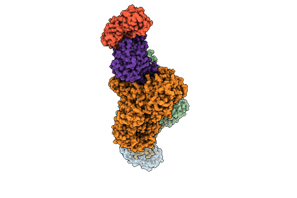
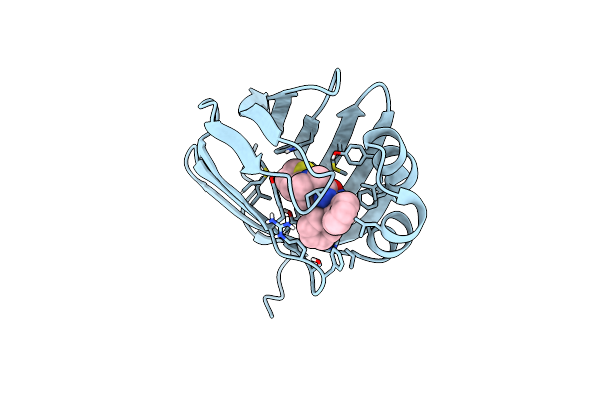
Organism: Severe acute respiratory syndrome coronavirus 2, Synthetic construct
Method: ELECTRON MICROSCOPY Release Date: 2025-03-12 Classification: VIRAL PROTEIN/RNA/INHIBITOR Ligands: ZN, A1AWQ, MN |
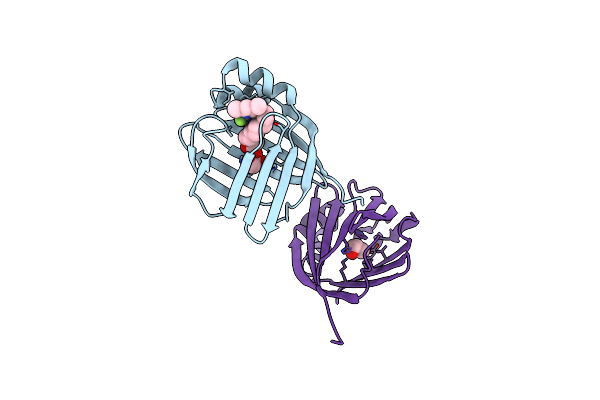
 |
Organism: Homo sapiens
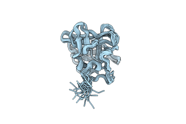
Method: ELECTRON MICROSCOPY Release Date: 2024-11-13 Classification: PROTEIN FIBRIL |
 |
X-Ray Structure Of The Peroxisomal Targeting Signal 1 (Pts1) Of Trypanosoma Cruzi Pex5 In Complex With The Pts1 Peptide
Organism: Trypanosoma cruzi
Method: X-RAY DIFFRACTION Release Date: 2024-10-02 Classification: TRANSPORT PROTEIN Ligands: EDO, PEG, MG, TRS |
 |
Crystal Structure Of The Apo Pex5 Peroxisomal Cargo Receptor From Trypanosoma Brucei
Organism: Trypanosoma brucei
Method: X-RAY DIFFRACTION Release Date: 2024-10-02 Classification: PEPTIDE BINDING PROTEIN |
 |
Cryo-Electron Microscopy Structure Of Glucose/Xylose Isomerase From Streptomyces Rubiginosus With Cobalt Ions In The Active Site
Organism: Streptomyces rubiginosus
Method: ELECTRON MICROSCOPY Release Date: 2024-10-02 Classification: METAL BINDING PROTEIN Ligands: CO |
 |
Cryo-Electron Microscopy Structure Of Glucose/Xylose Isomerase From Streptomyces Rubiginosus With Magnesium Ions In The Active Site
Organism: Streptomyces rubiginosus
Method: ELECTRON MICROSCOPY Release Date: 2024-10-02 Classification: METAL BINDING PROTEIN Ligands: MG |
 |
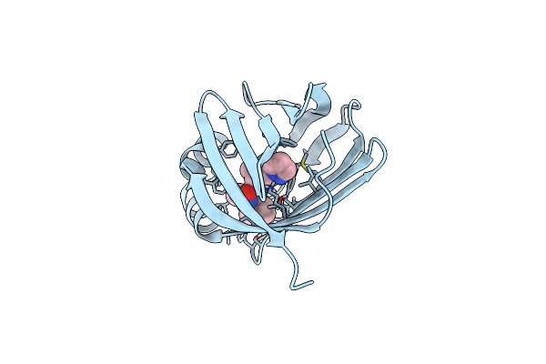
Crystal Structure Of Human Cellular Retinol Binding Protein 3 In Complex With C11 Topfluor Mg
Organism: Homo sapiens
Method: X-RAY DIFFRACTION Resolution:1.45 Å Release Date: 2024-04-17 Classification: LIPID BINDING PROTEIN Ligands: A1ALJ, GOL |
 |
Structure Of The Endothelial Monocyte Activating Polypeptide Ii (Emap Ii) In Solution
Organism: Homo sapiens
Method: SOLUTION NMR, SOLUTION SCATTERING Release Date: 2024-04-10 Classification: CYTOKINE |
 |
The Dbc1/Sirt1 Interaction Is Choreographed By Post-Translational Modification
Organism: Homo sapiens
Method: X-RAY DIFFRACTION Resolution:2.00 Å Release Date: 2024-03-27 Classification: GENE REGULATION |
 |
Organism: Mus musculus
Method: ELECTRON MICROSCOPY Release Date: 2023-10-11 Classification: TRANSPORT PROTEIN Ligands: NAG, U9L |
 |
Organism: Mus musculus
Method: ELECTRON MICROSCOPY Release Date: 2023-10-11 Classification: TRANSPORT PROTEIN Ligands: NAG, U9Q |
 |
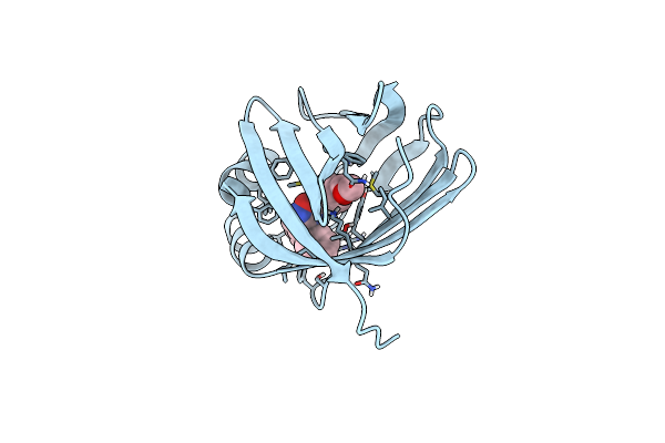
Crystal Structure Of Human Cellular Retinol Binding Protein 1 In Complex With Methyl({3-[1-(4-Methylphenyl)Cyclopentyl]-1,2,4-Oxadiazol-5-Yl}Methyl)[(1-Methylpyrazol-4-Yl)Methyl]Amine
Organism: Homo sapiens
Method: X-RAY DIFFRACTION Resolution:1.47 Å Release Date: 2023-10-11 Classification: Retinol-binding protein Ligands: ZCF, BTB |
 |
Crystal Structure Of Human Cellular Retinol Binding Protein 1 In Complex With N-Methyl-1-{3-[1-(4-Methylphenyl)Cyclopentyl]-1,2,4-Oxadiazol-5-Yl}-N-(2-Thienylmethyl)Methanamine
Organism: Homo sapiens
Method: X-RAY DIFFRACTION Resolution:1.13 Å Release Date: 2023-10-04 Classification: Retinol-binding protein Ligands: Z5H, BTB |
 |
Crystal Structure Of Human Cellular Retinol Binding Protein 1 In Complex With {[3-(Diphenylmethyl)-1,2,4-Oxadiazol-5-Yl]Methyl}(Methyl)[1-(Thiophen-2-Yl)Ethyl]Amine
Organism: Homo sapiens
Method: X-RAY DIFFRACTION Resolution:1.80 Å Release Date: 2023-10-04 Classification: Retinol-binding protein Ligands: ZA6 |
 |
Crystal Structure Of Human Cellular Retinol Binding Protein 1 In Complex With N-Ethyl-N-({3-[1-(4-Methylphenyl)Cyclopentyl]-1,2,4-Oxadiazol-5-Yl}Methyl)-2-(1H-Pyrazol-1-Yl)Ethanamine
Organism: Homo sapiens
Method: X-RAY DIFFRACTION Resolution:1.55 Å Release Date: 2023-10-04 Classification: retinol-binding protein Ligands: ZDF |
 |
Crystal Structure Of Human Cellular Retinol Binding Protein 1 In Complex With 1-{[3-(Diphenylmethyl)-1,2,4-Oxadiazol-5-Yl]Methyl}-4-(Methoxymethyl)Piperidine
Organism: Homo sapiens
Method: X-RAY DIFFRACTION Resolution:1.85 Å Release Date: 2023-10-04 Classification: Retinol-binding protein Ligands: ZDK |
 |
Crystal Structure Of Human Cellular Retinol Binding Protein 1 In Complex With 4-(Hydroxymethyl)-1-[(4-Methoxy-5,6,7,8-Tetrahydronaphthalen-1-Yl)Sulfonyl]Piperidin-4-Ol
Organism: Homo sapiens
Method: X-RAY DIFFRACTION Resolution:1.30 Å Release Date: 2023-10-04 Classification: Retinol-binding protein Ligands: ZE2, BTB |
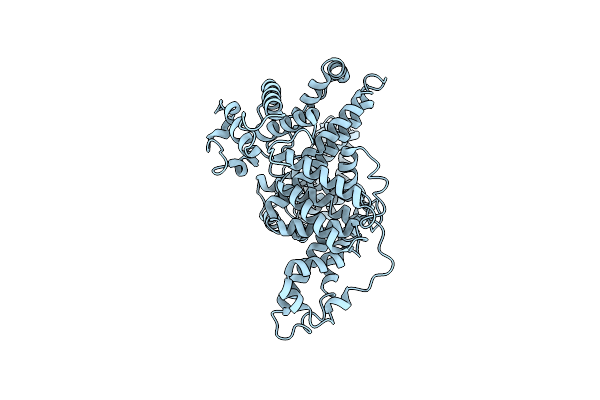 |
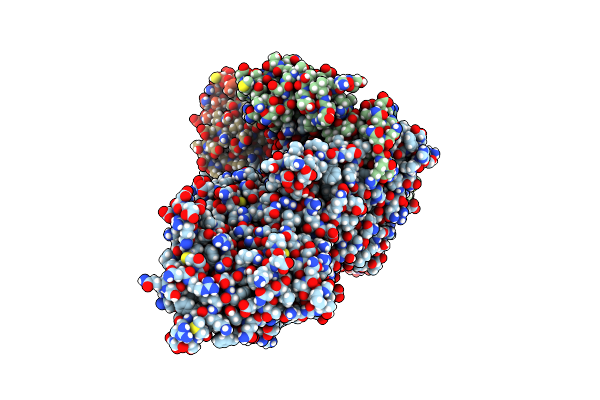
Structural Studies Of Human Serum Albumin Using Cryo-Em Up To 0.38 Nm Resolution
Organism: Homo sapiens
Method: ELECTRON MICROSCOPY Release Date: 2023-08-16 Classification: TRANSPORT PROTEIN |
 |
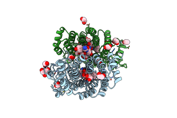
Crystal Structure Of E. Coli Fabi In Complex With Nad And (R,E)-3-(7-Amino-8-Oxo-6,7,8,9-Tetrahydro-5H-Pyrido[2,3-B]Azepin-3-Yl)-N-Methyl-N-((3-Methylbenzofuran-2-Yl)Methyl)Acrylamide
Organism: Escherichia coli str. k-12 substr. mg1655
Method: X-RAY DIFFRACTION Resolution:1.70 Å Release Date: 2023-02-15 Classification: ANTIBIOTIC Ligands: NAD, NQF |