
Search Count: 25
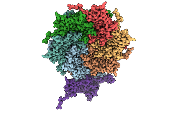 |
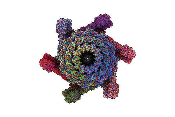
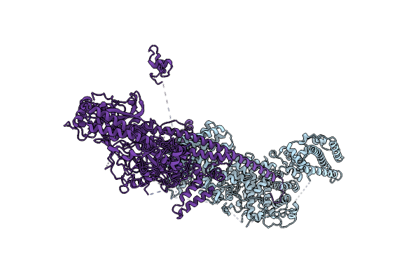
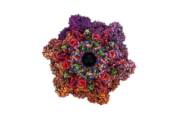
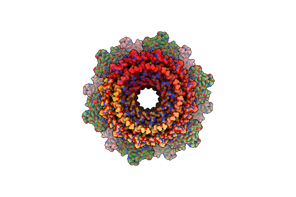
Organism: Pseudanabaena phage pan3
Method: ELECTRON MICROSCOPY Resolution:3.20 Å Release Date: 2025-11-26 Classification: VIRUS |
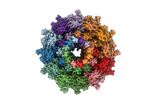 |
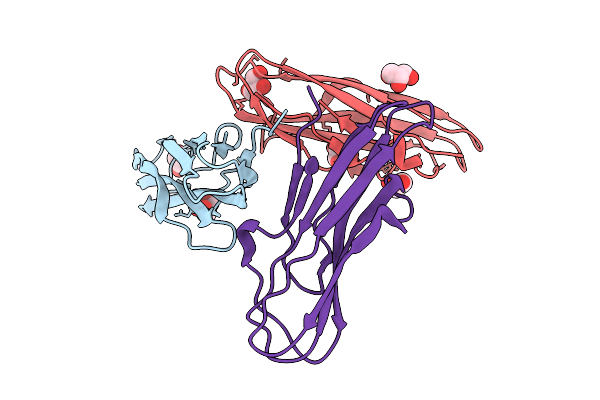
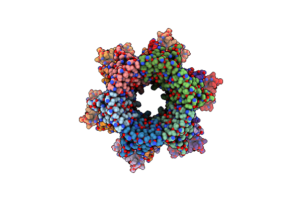
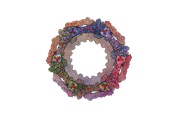
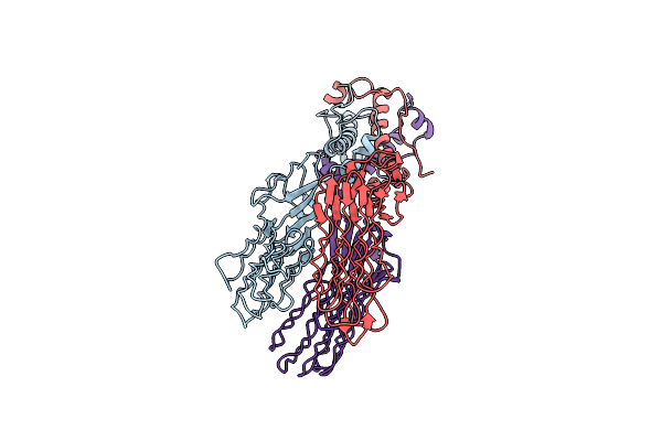
Organism: Pseudanabaena phage pan3
Method: ELECTRON MICROSCOPY Resolution:3.30 Å Release Date: 2025-11-26 Classification: VIRAL PROTEIN |
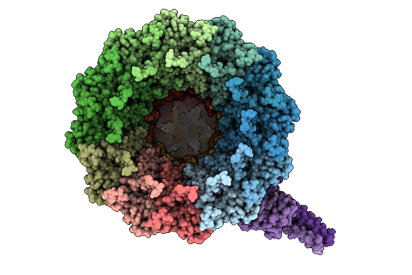 |
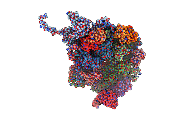
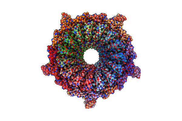
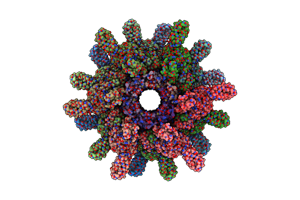
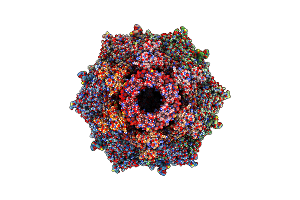
Organism: Pseudanabaena phage pan3
Method: ELECTRON MICROSCOPY Resolution:3.50 Å Release Date: 2025-11-26 Classification: VIRAL PROTEIN |
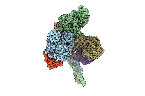 |
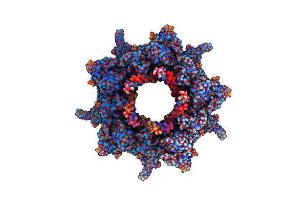
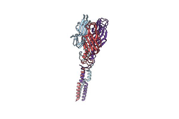
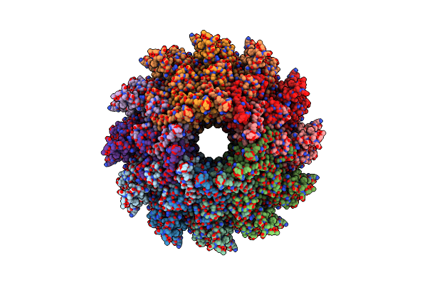
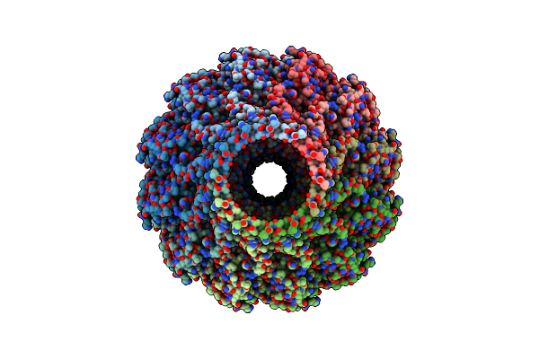
Organism: Pseudanabaena phage pan3
Method: ELECTRON MICROSCOPY Resolution:3.50 Å Release Date: 2025-11-26 Classification: VIRAL PROTEIN |
 |
Organism: Pseudanabaena phage pan3
Method: X-RAY DIFFRACTION Resolution:2.60 Å Release Date: 2025-11-26 Classification: VIRUS Ligands: IOD, GOL |
 |
Organism: Anabaena phage a-4l
Method: ELECTRON MICROSCOPY Resolution:2.80 Å Release Date: 2025-04-09 Classification: VIRUS |
 |
Organism: Anabaena phage a-4l
Method: ELECTRON MICROSCOPY Release Date: 2025-04-09 Classification: VIRAL PROTEIN |
 |
Organism: Anabaena phage a-4l
Method: ELECTRON MICROSCOPY Resolution:3.40 Å Release Date: 2025-04-09 Classification: VIRAL PROTEIN Ligands: ZN |
 |
Organism: Anabaena phage a-4l
Method: ELECTRON MICROSCOPY Resolution:3.60 Å Release Date: 2025-04-09 Classification: VIRAL PROTEIN |
 |
Organism: Unclassified caudoviricetes
Method: ELECTRON MICROSCOPY Release Date: 2024-04-17 Classification: VIRAL PROTEIN |
 |
Organism: Unclassified caudoviricetes
Method: ELECTRON MICROSCOPY Release Date: 2024-04-17 Classification: VIRAL PROTEIN |
 |
Organism: Unclassified caudoviricetes
Method: ELECTRON MICROSCOPY Release Date: 2024-04-10 Classification: VIRAL PROTEIN |
 |
Organism: Unclassified caudoviricetes
Method: ELECTRON MICROSCOPY Release Date: 2024-04-10 Classification: VIRAL PROTEIN |
 |
Organism: Unclassified caudoviricetes
Method: ELECTRON MICROSCOPY Release Date: 2024-04-10 Classification: VIRAL PROTEIN |
 |
Organism: Unclassified caudoviricetes
Method: ELECTRON MICROSCOPY Release Date: 2024-04-10 Classification: VIRAL PROTEIN |
 |
Organism: Uncultured cyanophage
Method: ELECTRON MICROSCOPY Release Date: 2024-04-10 Classification: VIRAL PROTEIN |
 |
Organism: Uncultured cyanophage
Method: ELECTRON MICROSCOPY Release Date: 2023-02-01 Classification: VIRAL PROTEIN |
 |
Organism: Uncultured cyanophage
Method: ELECTRON MICROSCOPY Release Date: 2023-01-18 Classification: VIRAL PROTEIN |
 |
Organism: Uncultured cyanophage
Method: ELECTRON MICROSCOPY Release Date: 2023-01-18 Classification: VIRAL PROTEIN |
 |
Organism: Uncultured cyanophage
Method: ELECTRON MICROSCOPY Release Date: 2023-01-18 Classification: VIRAL PROTEIN |