
Search Count: 16
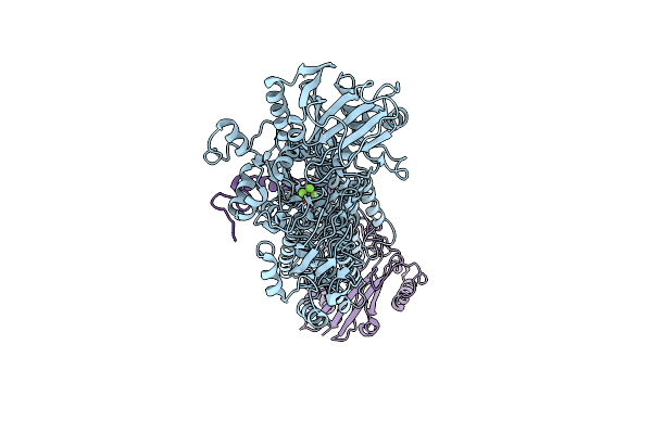 |
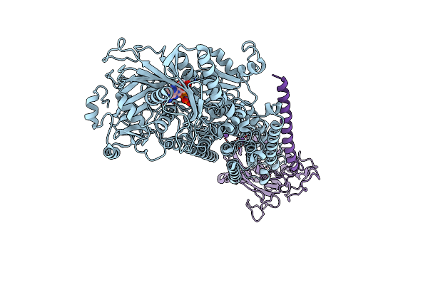
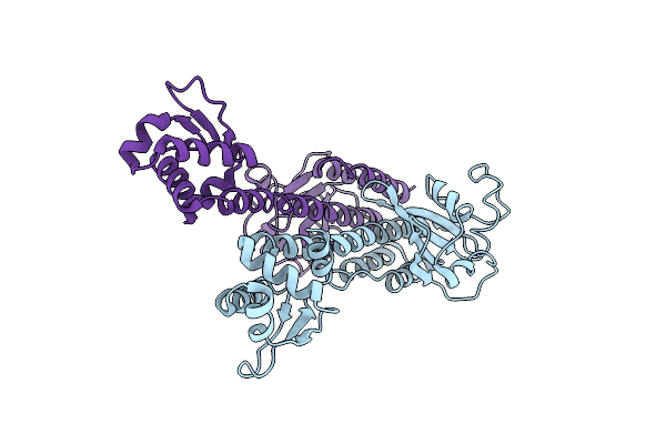
Cryo-Em Structure Of Na+,K+-Atpase Alpha2 From Artemia Salina In Cation-Free E2P Form
Organism: Artemia salina
Method: ELECTRON MICROSCOPY Release Date: 2023-11-29 Classification: TRANSPORT PROTEIN Ligands: ALF |
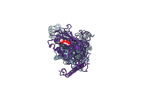 |
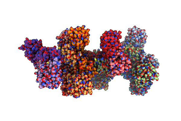
Organism: Rattus norvegicus
Method: X-RAY DIFFRACTION Resolution:3.30 Å Release Date: 2022-10-05 Classification: MEMBRANE PROTEIN Ligands: ALF, K, NAG |
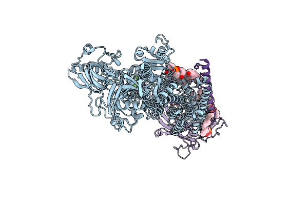 |
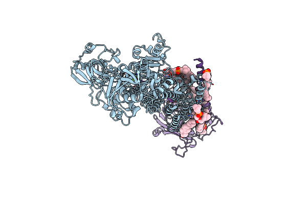
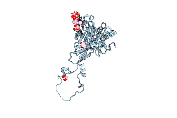
Cryo-Em Structure Of Non Gastric H,K-Atpase Alpha2 K794A In (K+)E2-Alf State
Organism: Rattus norvegicus
Method: ELECTRON MICROSCOPY Release Date: 2022-10-05 Classification: MEMBRANE PROTEIN Ligands: K, ALF, MG, CLR, PCW, NAG |
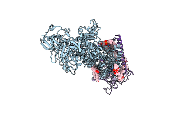 |
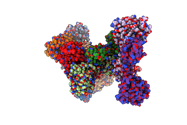
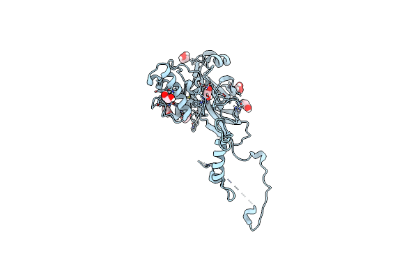
Cryo-Em Structure Of Non Gastric H,K-Atpase Alpha2 K794S In (2K+)E2-Alf State
Organism: Rattus norvegicus
Method: ELECTRON MICROSCOPY Release Date: 2022-10-05 Classification: MEMBRANE PROTEIN Ligands: ALF, MG, K, CLR, PCW, NAG |
 |
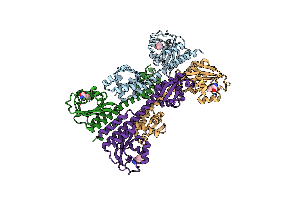
Cryo-Em Structure Of Non Gastric H,K-Atpase Alpha2 Spwc Mutant In 3Na+E1-Amppcpf State
Organism: Rattus norvegicus
Method: ELECTRON MICROSCOPY Release Date: 2022-10-05 Classification: MEMBRANE PROTEIN Ligands: ACP, NA, NAG |
 |
Cryo-Em Structure Of Non Gastric H,K-Atpase Alpha2 Spwc Mutant In (2K+)E2-Alf State
Organism: Rattus norvegicus
Method: ELECTRON MICROSCOPY Release Date: 2022-10-05 Classification: MEMBRANE PROTEIN Ligands: K, PCW, CLR, ALF, MG, NAG |
 |
Organism: Bacillus subtilis (strain 168)
Method: X-RAY DIFFRACTION Resolution:3.00 Å Release Date: 2017-01-11 Classification: TRANSCRIPTION Ligands: SO4 |
 |
Organism: Bacillus subtilis (strain 168)
Method: X-RAY DIFFRACTION Resolution:3.00 Å Release Date: 2017-01-11 Classification: TRANSCRIPTION Ligands: ILE |
 |
Organism: Bacillus subtilis (strain 168)
Method: X-RAY DIFFRACTION Resolution:3.71 Å Release Date: 2017-01-11 Classification: TRANSCRIPTION |
 |
Organism: Bacillus subtilis
Method: X-RAY DIFFRACTION Resolution:4.50 Å Release Date: 2017-01-11 Classification: TRANSCRIPTION |
 |
The Atomic Structure Of Anomala Cuprea Entomopoxvirus (Acepv) Fusolin Spindles
Organism: Anomala cuprea entomopoxvirus
Method: X-RAY DIFFRACTION Resolution:1.90 Å Release Date: 2015-04-08 Classification: VIRAL PROTEIN Ligands: EDO |
 |
The Atomic Structure Of Wiseana Spp Entomopoxvirus (Wsepv) Fusolin Spindles
Organism: Unidentified entomopoxvirus
Method: X-RAY DIFFRACTION Resolution:2.02 Å Release Date: 2015-04-08 Classification: VIRAL PROTEIN Ligands: ZN, EDO |
 |
Structural Basis For The Enhancement Of Virulence By Entomopoxvirus Fusolin And Its In Vivo Crystallization Into Viral Spindles
Organism: Unidentified entomopoxvirus
Method: X-RAY DIFFRACTION Resolution:1.90 Å Release Date: 2015-03-18 Classification: VIRAL PROTEIN Ligands: EDO |
 |
Structural Basis For The Enhancement Of Virulence By Entomopoxvirus Fusolin And Its In Vivo Crystallization Into Viral Spindles (Complex With Copper)
Organism: Entomopoxvirinae
Method: X-RAY DIFFRACTION Resolution:2.40 Å Release Date: 2015-03-18 Classification: VIRAL PROTEIN Ligands: CU |
 |
Structural Basis For The Enhancement Of Virulence By Entomopoxvirus Fusolin And Its In Vivo Crystallization Into Viral Spindles (Complex With Zinc)
Organism: Entomopoxvirinae
Method: X-RAY DIFFRACTION Resolution:2.41 Å Release Date: 2015-03-18 Classification: VIRAL PROTEIN Ligands: ZN |
 |
Organism: Rattus norvegicus
Method: X-RAY DIFFRACTION Resolution:2.05 Å Release Date: 2009-12-22 Classification: TRANSFERASE Ligands: MG, ZN, SZH |