
Search Count: 31
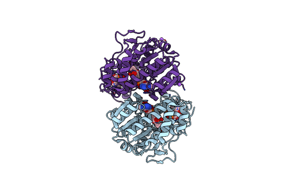 |
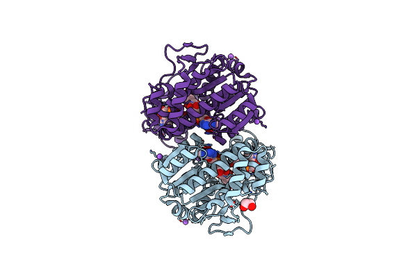
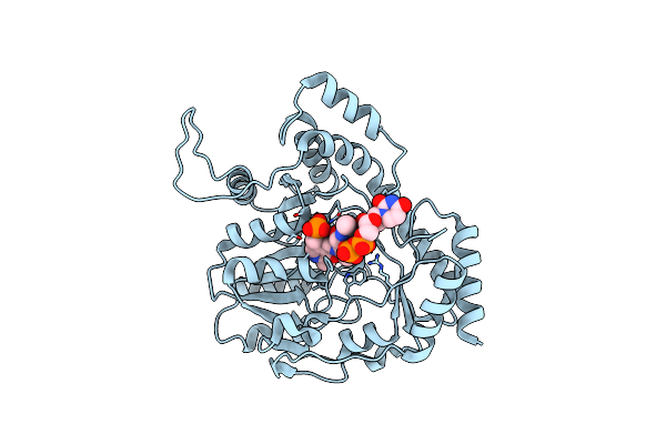
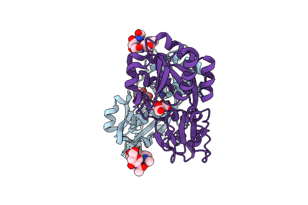
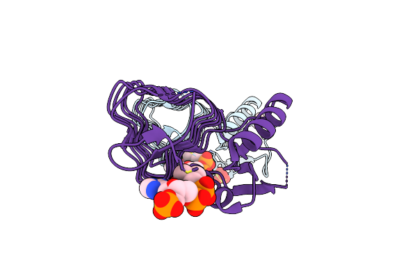
X-Ray Structure Of The Pglf Dehydratase From Campylobacter Jejuni In Complex With Udp And Nad(H)
Organism: Campylobacter jejuni
Method: X-RAY DIFFRACTION Resolution:2.00 Å Release Date: 2017-11-08 Classification: MEMBRANE PROTEIN Ligands: UDP, NAD, EDO, NA |
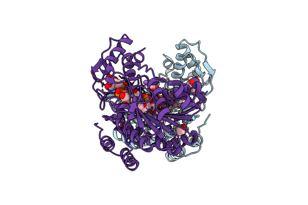 |
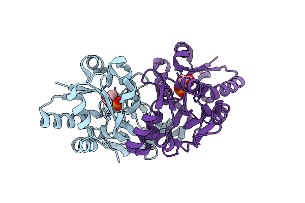
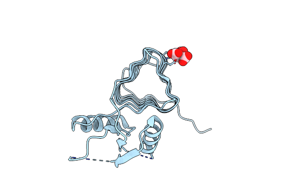
X-Ray Structure Of The Pglf Udp-N-Acetylglucosamine 4,6-Dehydratase From Campylobacterjejuni, D396N/K397A Variant In Complex With Udp-N-Acrtylglucosamine
Organism: Campylobacter jejuni
Method: X-RAY DIFFRACTION Resolution:1.80 Å Release Date: 2017-11-08 Classification: MEMBRANE PROTEIN Ligands: NAD, UD1, EDO, NA |
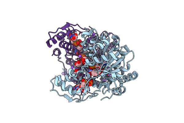 |
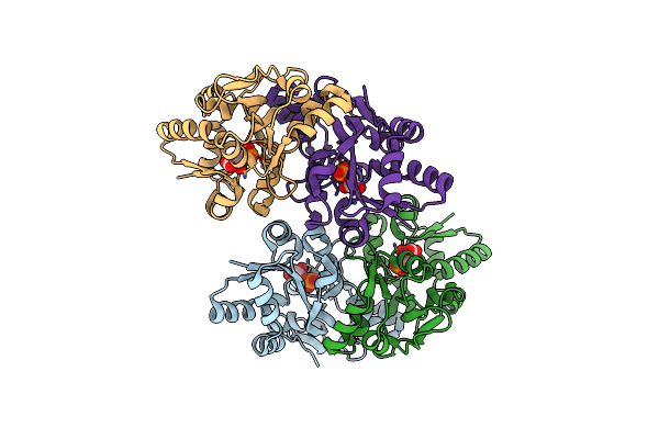
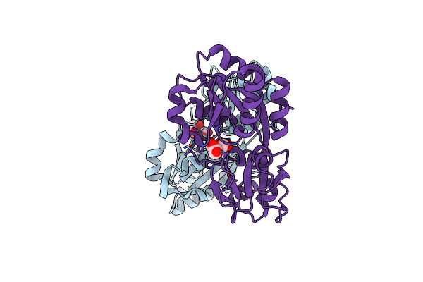
X-Ray Structure Of The Pglf 4,6-Dehydratase From Campylobacter Jejuni, T595S Variant, In Complex With Udp
Organism: Campylobacter jejuni
Method: X-RAY DIFFRACTION Resolution:1.60 Å Release Date: 2017-11-08 Classification: MEMBRANE PROTEIN Ligands: UDP, NAD, EDO, NA |
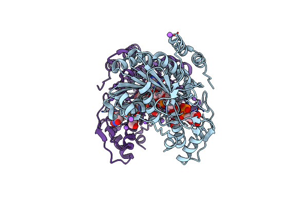 |
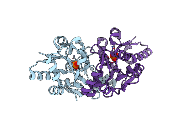
X-Ray Structure Of The Pglf 4,6-Dehydratase From Campylobacter Jejuni, Variant T395V, In Complex With Udp
Organism: Campylobacter jejuni
Method: X-RAY DIFFRACTION Resolution:1.60 Å Release Date: 2017-11-08 Classification: MEMBRANE PROTEIN Ligands: UDP, NAD, EDO, NA |
 |
X-Ray Structure Of The Pglf 4,5-Dehydratase From Campylobacter Jejuni, Variant M405Y, In Complex With Udp
Organism: Campylobacter jejuni
Method: X-RAY DIFFRACTION Resolution:1.60 Å Release Date: 2017-11-08 Classification: MEMBRANE PROTEIN Ligands: UDP, NAD, EDO, NA |
 |
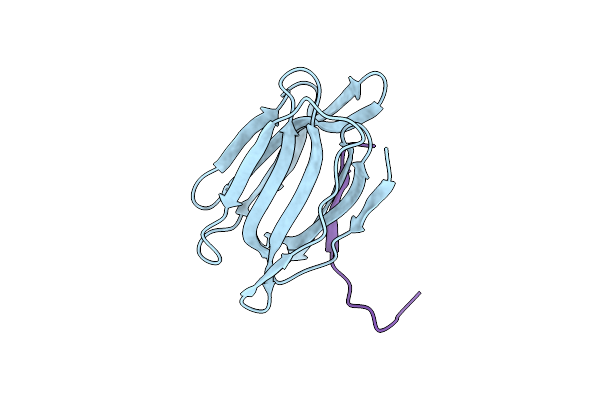
Organism: Wisteria floribunda
Method: X-RAY DIFFRACTION Resolution:2.33 Å Release Date: 2016-09-14 Classification: SUGAR BINDING PROTEIN Ligands: NGA, CA, MN |
 |
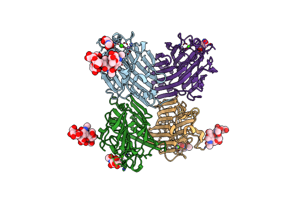
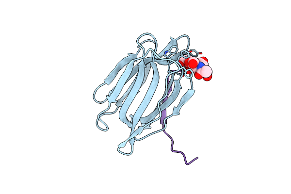
Wisteria Floribunda Lectin In Complex With Galnac(Beta1-4)Glcnac (Lacdinac) At Ph 8.5.
Organism: Wisteria floribunda
Method: X-RAY DIFFRACTION Resolution:1.80 Å Release Date: 2016-09-14 Classification: SUGAR BINDING PROTEIN Ligands: 6Y2, CA, MN, NAG |
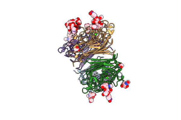 |
Wisteria Floribunda Lectin In Complex With Galnac(Beta1-4)Glcnac (Lacdinac) At Ph 6.5
Organism: Wisteria floribunda
Method: X-RAY DIFFRACTION Resolution:1.95 Å Release Date: 2016-09-14 Classification: SUGAR BINDING PROTEIN Ligands: CA, MN, ACT, 6Y2, NAG |
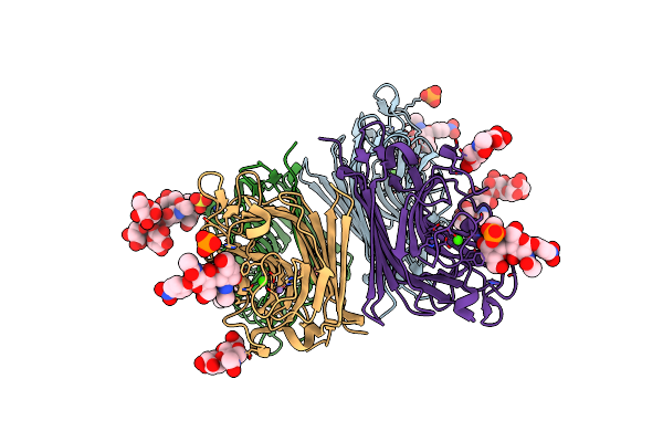 |
Wisteria Floribunda Lectin In Complex With Galnac(Beta1-4)Glcnac (Lacdinac) At Ph 4.2
Organism: Wisteria floribunda
Method: X-RAY DIFFRACTION Resolution:2.09 Å Release Date: 2016-09-14 Classification: sugar binding protein/inhibitor Ligands: 6Y2, MN, CA, PO4, SO4 |
 |
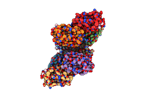
Organism: Campylobacter jejuni
Method: X-RAY DIFFRACTION Resolution:2.00 Å Release Date: 2015-07-29 Classification: TRANSFERASE Ligands: 4RA |
 |
Organism: Maclura pomifera
Method: X-RAY DIFFRACTION Resolution:2.25 Å Release Date: 2010-09-22 Classification: SUGAR BINDING PROTEIN |
 |
Crystal Structure Analysis Of Maclura Pomifera Agglutinin Complex With Gal-Beta-1,3-Galnac
Organism: Maclura pomifera
Method: X-RAY DIFFRACTION Resolution:1.55 Å Release Date: 2010-09-22 Classification: SUGAR BINDING PROTEIN |
 |
Crystal Structure Analysis Of Maclura Pomifera Agglutinin Complex With P-Nitrophenyl-Galnac
Organism: Maclura pomifera
Method: X-RAY DIFFRACTION Resolution:2.10 Å Release Date: 2010-09-22 Classification: SUGAR BINDING PROTEIN Ligands: LEC |
 |
Organism: Campylobacter jejuni
Method: X-RAY DIFFRACTION Resolution:2.00 Å Release Date: 2009-03-10 Classification: PROTEIN BINDING Ligands: FLC |
 |
Organism: Campylobacter jejuni
Method: X-RAY DIFFRACTION Resolution:1.70 Å Release Date: 2009-03-10 Classification: PROTEIN BINDING Ligands: PEQ |
 |
Organism: Campylobacter jejuni
Method: X-RAY DIFFRACTION Resolution:2.20 Å Release Date: 2009-03-10 Classification: TRANSPORT PROTEIN Ligands: 3PG |
 |
Organism: Campylobacter jejuni
Method: X-RAY DIFFRACTION Resolution:1.60 Å Release Date: 2009-03-10 Classification: TRANSPORT PROTEIN Ligands: PO4 |
 |
Organism: Campylobacter jejuni
Method: X-RAY DIFFRACTION Resolution:1.80 Å Release Date: 2008-01-29 Classification: TRANSFERASE Ligands: COA, SO4 |
 |
Organism: Campylobacter jejuni
Method: X-RAY DIFFRACTION Resolution:1.75 Å Release Date: 2008-01-22 Classification: TRANSFERASE Ligands: FLC |
 |
Organism: Campylobacter jejuni
Method: X-RAY DIFFRACTION Resolution:1.60 Å Release Date: 2007-05-01 Classification: periplasmic binding protein Ligands: FLC |