
Search Count: 26
 |
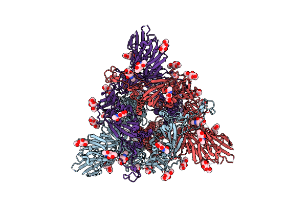
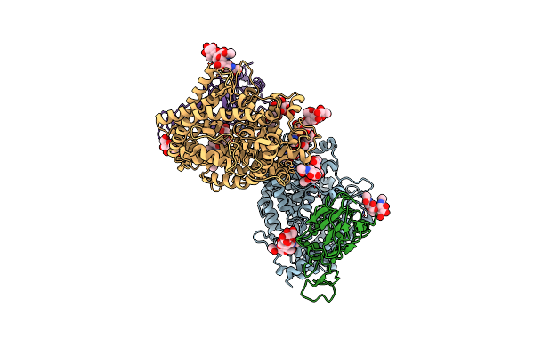
Structure Of The Sars-Cov-2 Eg.5.1 Spike Glycoprotein In Complex With Ace2 (1-Up State)
Organism: Severe acute respiratory syndrome coronavirus 2, Homo sapiens
Method: ELECTRON MICROSCOPY Release Date: 2024-05-01 Classification: VIRAL PROTEIN/PROTEIN BINDING Ligands: NAG |
 |
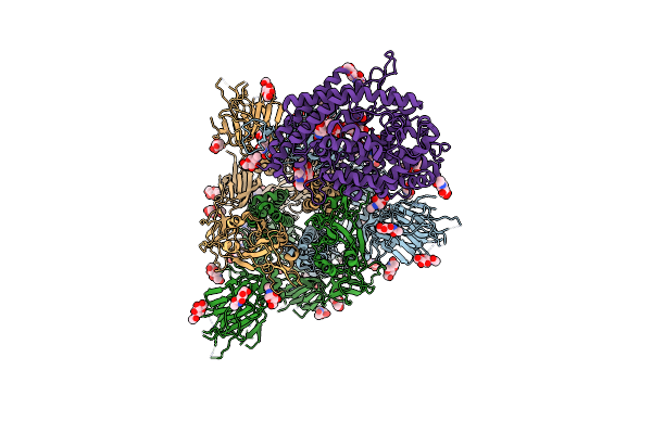
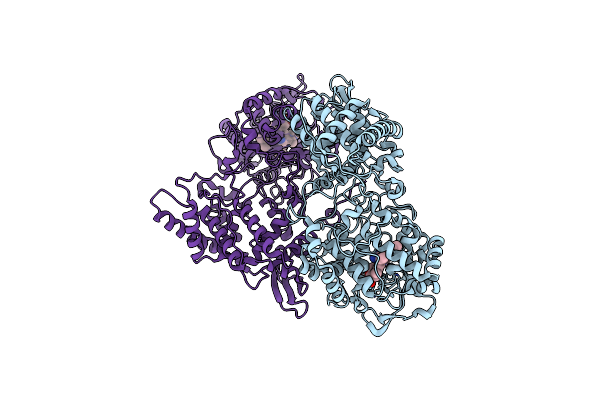
Organism: Homo sapiens, Severe acute respiratory syndrome coronavirus 2
Method: ELECTRON MICROSCOPY Release Date: 2024-05-01 Classification: VIRAL PROTEIN/PROTEIN BINDING Ligands: NAG |
 |
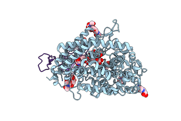
Organism: Severe acute respiratory syndrome coronavirus 2
Method: ELECTRON MICROSCOPY Release Date: 2024-04-24 Classification: VIRAL PROTEIN Ligands: NAG |
 |
Organism: Severe acute respiratory syndrome coronavirus 2
Method: ELECTRON MICROSCOPY Release Date: 2024-04-24 Classification: VIRAL PROTEIN Ligands: NAG |
 |
Organism: Severe acute respiratory syndrome coronavirus 2
Method: ELECTRON MICROSCOPY Release Date: 2024-01-03 Classification: VIRAL PROTEIN Ligands: NAG |
 |
Organism: Severe acute respiratory syndrome coronavirus 2
Method: ELECTRON MICROSCOPY Release Date: 2024-01-03 Classification: VIRAL PROTEIN Ligands: NAG |
 |
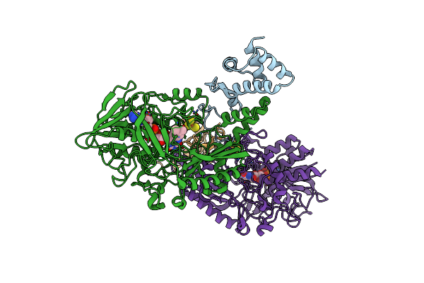
Structure Of Sars-Cov-2 Xbb.1.5 Spike Glycoprotein In Complex With Ace2 (1-Up State)
Organism: Severe acute respiratory syndrome coronavirus 2, Homo sapiens
Method: ELECTRON MICROSCOPY Release Date: 2024-01-03 Classification: VIRAL PROTEIN/PROTEIN BINDING Ligands: NAG |
 |
Structure Of Sars-Cov-2 Xbb.1.5 Spike Glycoprotein In Complex With Ace2 (2-Up State)
Organism: Severe acute respiratory syndrome coronavirus 2, Homo sapiens
Method: ELECTRON MICROSCOPY Release Date: 2024-01-03 Classification: VIRAL PROTEIN/PROTEIN BINDING Ligands: NAG |
 |
Organism: Severe acute respiratory syndrome coronavirus 2, Homo sapiens
Method: ELECTRON MICROSCOPY Release Date: 2024-01-03 Classification: VIRAL PROTEIN/PROTEIN BINDING Ligands: NAG |
 |
Organism: Severe acute respiratory syndrome coronavirus 2
Method: ELECTRON MICROSCOPY Release Date: 2023-05-24 Classification: VIRAL PROTEIN Ligands: NAG |
 |
Organism: Severe acute respiratory syndrome coronavirus 2
Method: ELECTRON MICROSCOPY Release Date: 2023-05-24 Classification: VIRAL PROTEIN Ligands: NAG |
 |
Structure Of Sars-Cov-2 Xbb.1 Spike Glycoprotein In Complex With Ace2 (1-Up State)
Organism: Severe acute respiratory syndrome coronavirus 2, Homo sapiens
Method: ELECTRON MICROSCOPY Release Date: 2023-05-24 Classification: VIRAL PROTEIN/PROTEIN BINDING Ligands: NAG |
 |
Organism: Homo sapiens, Severe acute respiratory syndrome coronavirus 2
Method: ELECTRON MICROSCOPY Release Date: 2023-05-24 Classification: VIRAL PROTEIN/PROTEIN BINDING Ligands: NAG |
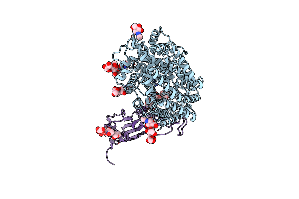 |
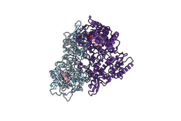
Crystal Structure Of The Receptor Binding Domain Of Sars-Cov-2 Omicron Bq.1.1 Variant Spike Protein In Complex With Its Receptor Ace2
Organism: Homo sapiens, Severe acute respiratory syndrome coronavirus 2
Method: X-RAY DIFFRACTION Resolution:2.78 Å Release Date: 2023-05-17 Classification: VIRAL PROTEIN Ligands: ZN, NAG |
 |
Organism: Severe acute respiratory syndrome coronavirus 2
Method: ELECTRON MICROSCOPY Release Date: 2022-10-26 Classification: VIRAL PROTEIN Ligands: NAG |
 |
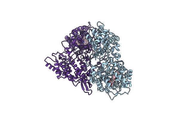
Crystal Structure Of The Receptor Binding Domain Of Sars-Cov-2 Omicron Ba.4/5 Variant Spike Protein In Complex With Its Receptor Ace2
Organism: Homo sapiens, Severe acute respiratory syndrome coronavirus 2
Method: X-RAY DIFFRACTION Resolution:3.36 Å Release Date: 2022-09-28 Classification: VIRAL PROTEIN Ligands: ZN, NAG |
 |
Crystal Structure Of Gamma-Alpha Subunit Complex From Burkholderia Cepacia Fad Glucose Dehydrogenase
Organism: Burkholderia cepacia
Method: X-RAY DIFFRACTION Resolution:2.60 Å Release Date: 2019-06-19 Classification: SIGNALING PROTEIN/OXIDOREDUCTASE Ligands: FAD, F3S |
 |
Crystal Structure Analysis Of The Ser305Thr Variants Of Katg From Haloarcula Marismortui
Organism: Haloarcula marismortui
Method: X-RAY DIFFRACTION Resolution:2.35 Å Release Date: 2012-12-05 Classification: OXIDOREDUCTASE Ligands: HEM |
 |
Crystal Structure Analysis Of The Arg409Leu Variants Of Katg From Haloarcula Marismortui
Organism: Haloarcula marismortui
Method: X-RAY DIFFRACTION Resolution:1.73 Å Release Date: 2012-12-05 Classification: OXIDOREDUCTASE Ligands: HEM |
 |
Crystal Structure Analysis Of The Cyanide Arg409Leu Variant Katg From Haloarcula Marismortui
Organism: Haloarcula marismortui
Method: X-RAY DIFFRACTION Resolution:1.70 Å Release Date: 2012-12-05 Classification: OXIDOREDUCTASE Ligands: HEM, CYN |