
Search Count: 37
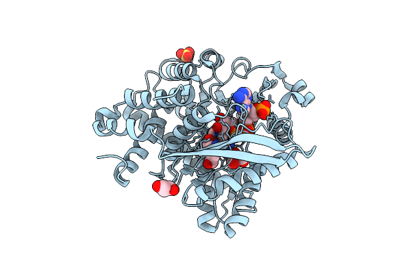 |
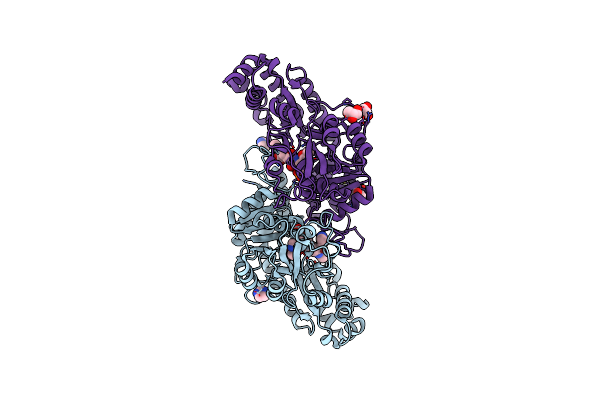
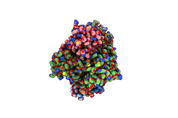
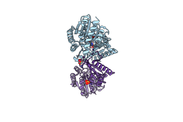
Crystal Structure Of Nadp-Specific Glutamate Dehydrogenase Gdh1 From Schizosaccharomyces Pombe In Complex With Alpha-Iminoglutarate And Nadp+
Organism: Schizosaccharomyces pombe
Method: X-RAY DIFFRACTION Release Date: 2025-07-30 Classification: OXIDOREDUCTASE Ligands: NAP, SO4, 2IT, GOL |
 |
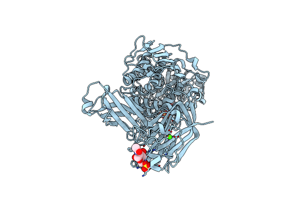
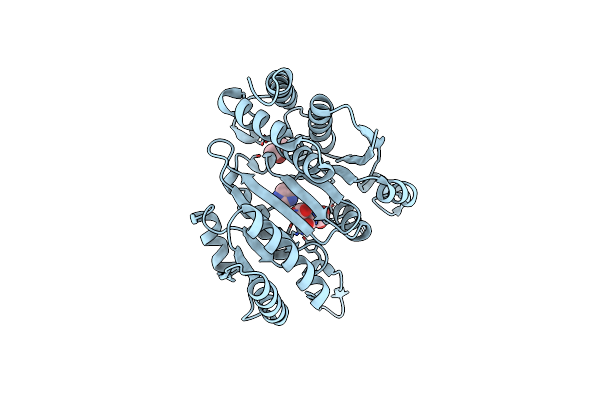
Organism: Scytonema tolypothrichoides vb-61278
Method: X-RAY DIFFRACTION Resolution:1.76 Å Release Date: 2025-04-09 Classification: BIOSYNTHETIC PROTEIN Ligands: SO4 |
 |
Crystal Structure Of A Heterooligomeric Aminotransferase From Serratia Sp. Atcc 39006
Organism: Serratia sp. atcc 39006
Method: X-RAY DIFFRACTION Resolution:2.84 Å Release Date: 2024-12-18 Classification: TRANSFERASE Ligands: SO4 |
 |
Crystal Structure Of A Heterooligomeric Aminotransferase From Serratia Sp. Atcc 39006, Pmp-Bound Form
Organism: Serratia sp. atcc 39006
Method: X-RAY DIFFRACTION Resolution:2.83 Å Release Date: 2024-12-18 Classification: TRANSFERASE Ligands: PMP |
 |
Crystal Structure Of A Heterooligomeric Aminotransferase From Serratia Sp. Atcc 39006, Ppe-Bound Form
Organism: Serratia sp. atcc 39006
Method: X-RAY DIFFRACTION Resolution:2.31 Å Release Date: 2024-12-18 Classification: TRANSFERASE Ligands: PGU |
 |
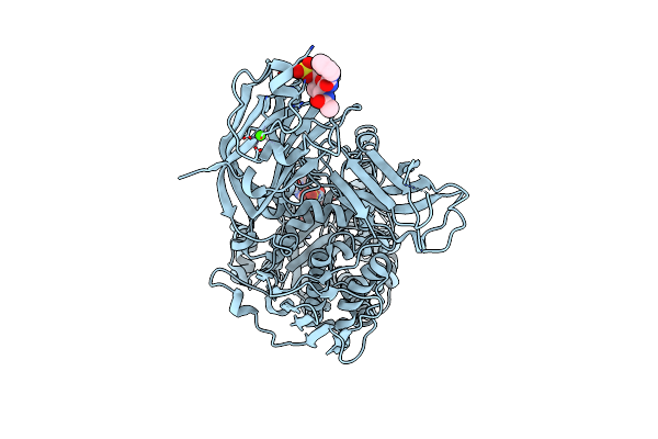
Organism: Streptomyces ficellus
Method: X-RAY DIFFRACTION Resolution:1.80 Å Release Date: 2023-02-22 Classification: BIOSYNTHETIC PROTEIN Ligands: GOL, IMD |
 |
Organism: Streptomyces ficellus
Method: X-RAY DIFFRACTION Resolution:1.93 Å Release Date: 2023-02-22 Classification: BIOSYNTHETIC PROTEIN Ligands: GOL, IMD, PLP |
 |
Crystal Structure Of Fic25 Complexed With Plp-(5S,6S)-N2-Acetyl-Dadh Adduct From Streptomyces Ficellus
Organism: Streptomyces ficellus
Method: X-RAY DIFFRACTION Resolution:1.82 Å Release Date: 2023-02-22 Classification: BIOSYNTHETIC PROTEIN Ligands: GOL, IMD, KKX |
 |
6-Sulfo-Beta-D-N-Acetylglucosaminidase From Bifidobacterium Bifidum In Complex With Glcnac-6S
Organism: Bifidobacterium bifidum jcm 1254
Method: X-RAY DIFFRACTION Resolution:1.65 Å Release Date: 2022-12-28 Classification: HYDROLASE Ligands: NGS, NGY, CA |
 |
6-Sulfo-Beta-D-N-Acetylglucosaminidase From Bifidobacterium Bifidum In Complex With Pugnac-6S
Organism: Bifidobacterium bifidum jcm 1254
Method: X-RAY DIFFRACTION Resolution:2.23 Å Release Date: 2022-12-28 Classification: HYDROLASE Ligands: 8R9, CA |
 |
Organism: Vibrio cholerae
Method: X-RAY DIFFRACTION Resolution:2.32 Å Release Date: 2022-11-09 Classification: CELL ADHESION Ligands: SO4 |
 |
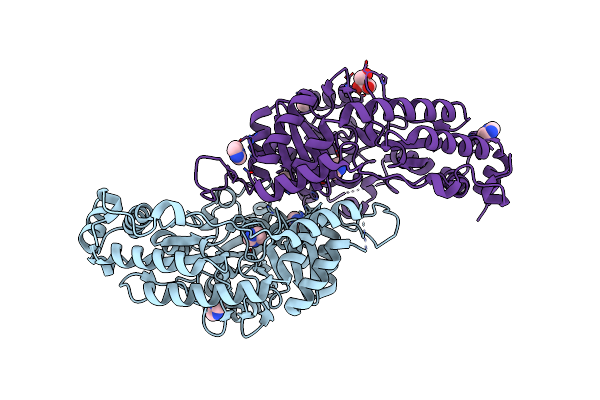
Crystal Structure Of Minor Pilin Tcpb From Vibrio Cholerae Complexed With N-Terminal Peptide Fragment Of Tcpf
Organism: Vibrio cholerae
Method: X-RAY DIFFRACTION Resolution:2.30 Å Release Date: 2022-11-09 Classification: CELL ADHESION Ligands: CA, CL, 1PE |
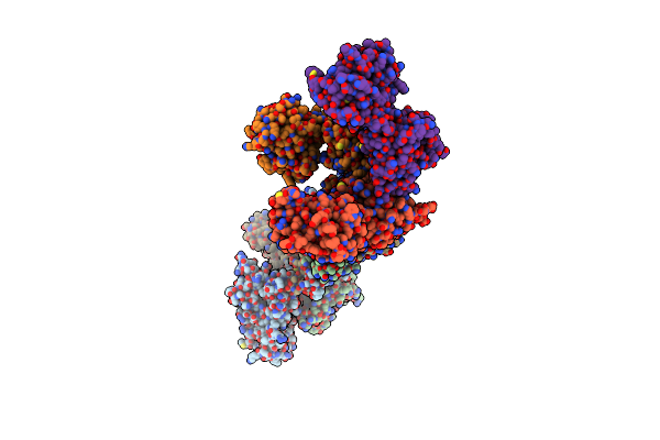 |
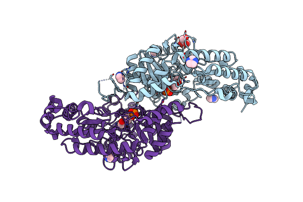
Crystal Structure Of Minor Pilin Tcpb From Vibrio Cholerae Complexed With Secreted Protein Tcpf
Organism: Vibrio cholerae
Method: X-RAY DIFFRACTION Resolution:4.05 Å Release Date: 2022-11-09 Classification: CELL ADHESION |
 |
Organism: Streptomyces sahachiroi
Method: X-RAY DIFFRACTION Resolution:1.75 Å Release Date: 2022-09-07 Classification: BIOSYNTHETIC PROTEIN Ligands: PG4, MG |
 |
Crystal Structure Of Aziu3/U2 Complexed With (5S,6S)-O7-Sulfo Dadh From Streptomyces Sahachiroi
Organism: Streptomyces sahachiroi
Method: X-RAY DIFFRACTION Resolution:1.80 Å Release Date: 2022-09-07 Classification: BIOSYNTHETIC PROTEIN Ligands: PGE, P4G, 6OI, MG |
 |
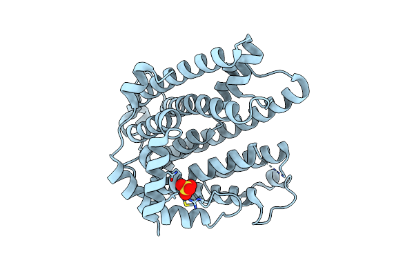
Organism: Thermus thermophilus (strain hb27 / atcc baa-163 / dsm 7039)
Method: X-RAY DIFFRACTION Resolution:2.39 Å Release Date: 2017-09-13 Classification: HYDROLASE Ligands: LYS, SO4 |
 |
Organism: Streptomyces melanosporofaciens
Method: X-RAY DIFFRACTION Resolution:1.80 Å Release Date: 2017-06-07 Classification: BIOSYNTHETIC PROTEIN Ligands: FMT |
 |
Crystal Structure Of Cotb2 (Ggspp/Mg2+-Bound Form) From Streptomyces Melanosporofaciens
Organism: Streptomyces melanosporofaciens
Method: X-RAY DIFFRACTION Resolution:1.80 Å Release Date: 2017-06-07 Classification: BIOSYNTHETIC PROTEIN Ligands: GGS, MG |
 |
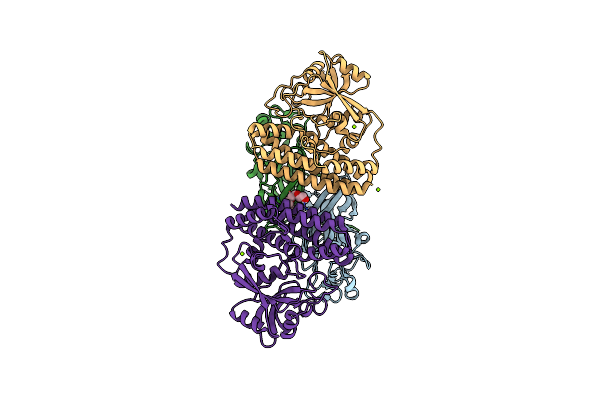
Crystal Structure Of Beta-Decarboxylating Dehydrogenase (Tk0280) From Thermococcus Kodakarensis (Apo Form)
Organism: Thermococcus kodakarensis (strain atcc baa-918 / jcm 12380 / kod1)
Method: X-RAY DIFFRACTION Resolution:1.70 Å Release Date: 2016-12-07 Classification: OXIDOREDUCTASE Ligands: IMD, NA |
 |
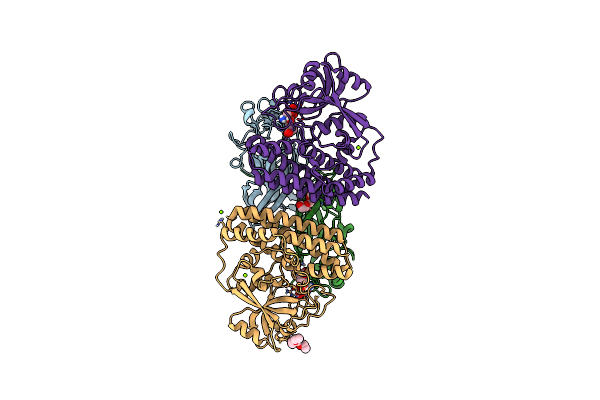
Crystal Structure Of Beta-Decarboxylating Dehydrogenase (Tk0280) From Thermococcus Kodakarensis Complexed With Mn And Homoisocitrate
Organism: Thermococcus kodakarensis (strain atcc baa-918 / jcm 12380 / kod1)
Method: X-RAY DIFFRACTION Resolution:2.64 Å Release Date: 2016-12-07 Classification: OXIDOREDUCTASE Ligands: 48Y, MN, IMD, IPA |