
Search Count: 91
 |
Organism: Severe acute respiratory syndrome coronavirus 2, Synthetic construct
Method: X-RAY DIFFRACTION Release Date: 2025-05-28 Classification: VIRAL PROTEIN |
 |
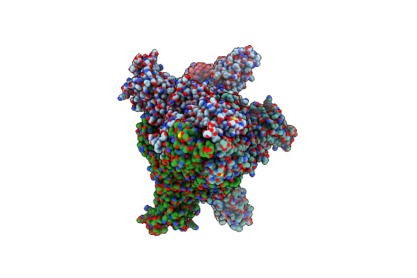
Organism: Severe acute respiratory syndrome coronavirus 2, Enterobacteria phage t4, Synthetic construct, Homo sapiens
Method: ELECTRON MICROSCOPY Release Date: 2025-01-22 Classification: VIRAL PROTEIN |
 |
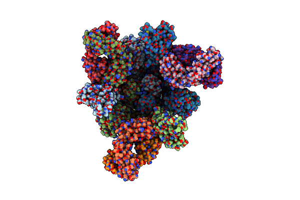
Organism: Severe acute respiratory syndrome coronavirus 2, Enterobacteria phage t4, Synthetic construct, Homo sapiens
Method: ELECTRON MICROSCOPY Release Date: 2025-01-22 Classification: VIRAL PROTEIN |
 |
Organism: Severe acute respiratory syndrome coronavirus 2, Enterobacteria phage t4, Synthetic construct, Homo sapiens
Method: ELECTRON MICROSCOPY Release Date: 2025-01-22 Classification: VIRAL PROTEIN |
 |
Organism: Severe acute respiratory syndrome coronavirus 2, Tequatrovirus t4, Synthetic construct, Homo sapiens
Method: ELECTRON MICROSCOPY Release Date: 2025-01-22 Classification: VIRAL PROTEIN |
 |
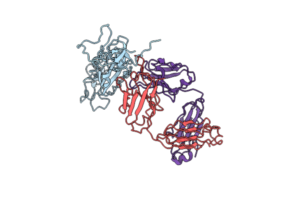
State 2 Of Sars-Cov-2 Xbb Variant Spike Protein Trimer Complexed With Antibody Pw5-5
Organism: Severe acute respiratory syndrome coronavirus 2, Homo sapiens
Method: ELECTRON MICROSCOPY Release Date: 2024-09-04 Classification: VIRAL PROTEIN/IMMUNE SYSTEM |
 |
Organism: Severe acute respiratory syndrome coronavirus 2, Homo sapiens
Method: ELECTRON MICROSCOPY Release Date: 2024-08-14 Classification: VIRAL PROTEIN/IMMUNE SYSTEM |
 |
The Local Refined Map Of Sars-Cov-2 Xbb Variant Spike Protein Complexed With Antibody Pw5-535
Organism: Homo sapiens, Severe acute respiratory syndrome coronavirus 2
Method: ELECTRON MICROSCOPY Release Date: 2024-08-14 Classification: IMMUNE SYSTEM/VIRAL PROTEIN |
 |
Organism: Homo sapiens, Severe acute respiratory syndrome coronavirus 2
Method: ELECTRON MICROSCOPY Release Date: 2024-08-14 Classification: IMMUNE SYSTEM/VIRAL PROTEIN |
 |
The Local Refined Map Of Sars-Cov Spike Protein Complexed With Antibody Pw5-5
Organism: Severe acute respiratory syndrome coronavirus 2, Homo sapiens
Method: ELECTRON MICROSCOPY Release Date: 2024-08-14 Classification: VIRAL PROTEIN/IMMUNE SYSTEM |
 |
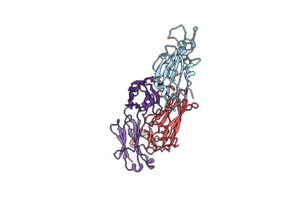
Monomer State Of Sars-Cov-2 Xbb Variant Spike Protein Trimer Complexed With Antibody Pw5-5
Organism: Homo sapiens, Severe acute respiratory syndrome coronavirus 2
Method: ELECTRON MICROSCOPY Release Date: 2024-08-14 Classification: IMMUNE SYSTEM/VIRAL PROTEIN |
 |
Organism: Homo sapiens, Severe acute respiratory syndrome coronavirus 2
Method: ELECTRON MICROSCOPY Release Date: 2024-08-14 Classification: IMMUNE SYSTEM/VIRAL PROTEIN |
 |
Organism: Severe acute respiratory syndrome coronavirus 2, Homo sapiens
Method: ELECTRON MICROSCOPY Release Date: 2024-08-14 Classification: VIRAL PROTEIN/IMMUNE SYSTEM |
 |
The Local Refined Map Of Sars-Cov-2 Omicron Ba.1 Spike Complexed With Antibody Pw5-570
Organism: Homo sapiens, Severe acute respiratory syndrome coronavirus 2
Method: ELECTRON MICROSCOPY Release Date: 2024-08-14 Classification: IMMUNE SYSTEM/VIRAL PROTEIN |
 |
State 1 Of Sars-Cov-2 Xbb Variant Spike Protein Trimer Complexed With Antibody Pw5-5
Organism: Severe acute respiratory syndrome coronavirus 2, Homo sapiens
Method: ELECTRON MICROSCOPY Release Date: 2024-08-14 Classification: VIRAL PROTEIN/IMMUNE SYSTEM |
 |
Structure Of Sars-Cov-2 Xbb Variant Spike Protein Complexed With Broadly Neutralizing Antibody Pw5-535
Organism: Severe acute respiratory syndrome coronavirus 2, Homo sapiens
Method: ELECTRON MICROSCOPY Release Date: 2024-08-14 Classification: IMMUNE SYSTEM/VIRAL PROTEIN |
 |
Organism: Bos taurus
Method: X-RAY DIFFRACTION Resolution:2.38 Å Release Date: 2023-09-27 Classification: MEMBRANE PROTEIN Ligands: CU, MG, NA, PGV, HEA, OH, CUA, TGL, PSC, CHD, PEK, CDL, DMU, ZN, SAC |
 |
Xfel Structure Of Mycobacterium Tuberculosis Beta Lactamase Microcrystals Mixed With Sulbactam For 3 Ms
Organism: Mycobacterium tuberculosis str. beijing/w bt1
Method: X-RAY DIFFRACTION Resolution:2.62 Å Release Date: 2023-09-20 Classification: HYDROLASE/Inhibitor Ligands: PO4 |
 |
Xfel Structure Of Mycobacterium Tuberculosis Beta Lactamase Microcrystals Mixed With Sulbactam For 6 Ms
Organism: Mycobacterium tuberculosis
Method: X-RAY DIFFRACTION Resolution:2.75 Å Release Date: 2023-09-20 Classification: HYDROLASE/Inhibitor Ligands: PO4 |
 |
Organism: Mycobacterium tuberculosis
Method: X-RAY DIFFRACTION Resolution:2.20 Å Release Date: 2023-09-20 Classification: HYDROLASE/Inhibitor Ligands: PO4 |