
Search Count: 315
 |
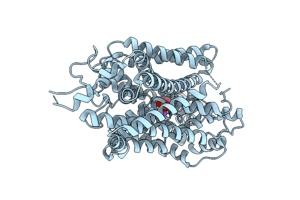
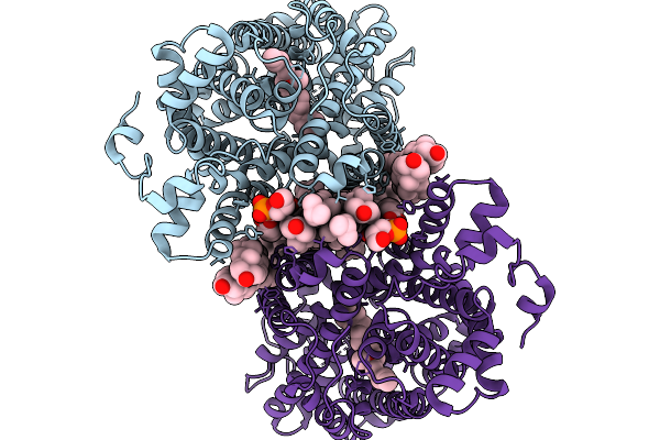
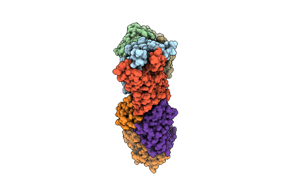
Cryo-Em Structure Of Conivaptan-Bound Human Vasopressin V2 Receptor Complex With Fab
Organism: Homo sapiens, Escherichia coli
Method: ELECTRON MICROSCOPY Release Date: 2025-12-10 Classification: MEMBRANE PROTEIN/IMMUNE SYSTEM Ligands: A1ECE |
 |
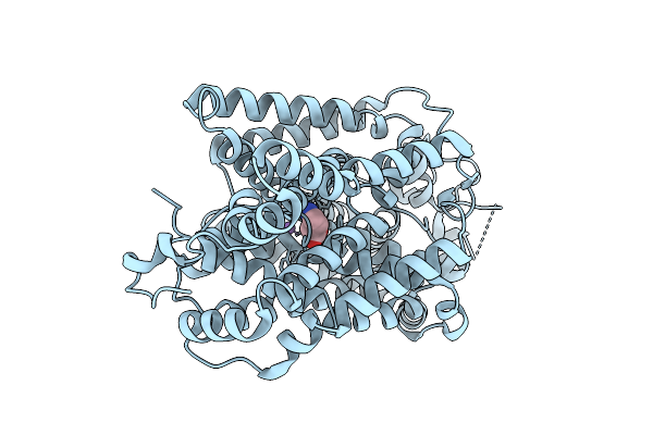
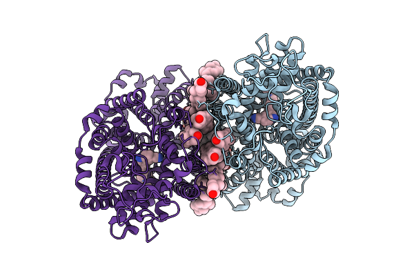
Cryo-Em Structure Of Tolvaptan-Bound Human Vasopressin V2 Receptor Complex With Fab
Organism: Homo sapiens, Escherichia coli
Method: ELECTRON MICROSCOPY Release Date: 2025-12-10 Classification: MEMBRANE PROTEIN/IMMUNE SYSTEM Ligands: A1ECF |
 |
Organism: Homo sapiens
Method: ELECTRON MICROSCOPY Release Date: 2025-12-03 Classification: MEMBRANE PROTEIN Ligands: CL |
 |
Organism: Homo sapiens
Method: ELECTRON MICROSCOPY Release Date: 2025-12-03 Classification: MEMBRANE PROTEIN Ligands: TAU, CL, NA |
 |
Organism: Homo sapiens
Method: ELECTRON MICROSCOPY Release Date: 2025-12-03 Classification: MEMBRANE PROTEIN Ligands: BAL, CL, NA |
 |
Organism: Homo sapiens
Method: ELECTRON MICROSCOPY Release Date: 2025-12-03 Classification: MEMBRANE PROTEIN Ligands: ABU, CL, NA |
 |
Organism: Homo sapiens
Method: ELECTRON MICROSCOPY Release Date: 2025-12-03 Classification: MEMBRANE PROTEIN Ligands: 3S5, CL |
 |
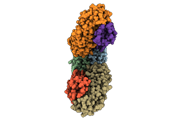
Cryo-Em Structure Of Lipid-Mediated Dimer Of Human Norepinephrine Transporter Net In The Presence Of Vanoxerine In An Inward-Open State At Resolution Of 2.52 Angstrom
Organism: Homo sapiens
Method: ELECTRON MICROSCOPY Release Date: 2025-11-19 Classification: TRANSPORT PROTEIN Ligands: CLR, A1D5S, A1EFM |
 |
Cryo-Em Structure Of Lipid-Mediated Human Norepinephrine Transporter Net In The Presence Of Levomilnacipran In An Inward-Open State At Resolution Of 2.46 Angstrom.
Organism: Homo sapiens
Method: ELECTRON MICROSCOPY Release Date: 2025-11-19 Classification: TRANSPORT PROTEIN Ligands: F0F, CLR, A1EFM |
 |
Cryo-Em Structure Of Lipid-Mediated Dimer Of Human Norepinephrine Transporter Net In The Presence Of The F3288-0031 In An Inward-Open State At Resolution Of 3.1 Angstrom
Organism: Homo sapiens
Method: ELECTRON MICROSCOPY Release Date: 2025-11-19 Classification: TRANSPORT PROTEIN Ligands: A1EFR, CLR, A1EFM |
 |
Cryo-Em Structure Of Lipid-Mediated Dimer Of Human Norepinephrine Transporter Net In The Presence Of The Antidepressant Vilazodone In An Inward-Open State At Resolution Of 2.44 Angstrom.
Organism: Homo sapiens
Method: ELECTRON MICROSCOPY Release Date: 2025-11-12 Classification: PROTON TRANSPORT Ligands: YG7, CLR, A1EFM |
 |
Organism: Sars-cov-2 pseudovirus, Homo sapiens
Method: ELECTRON MICROSCOPY Release Date: 2025-10-29 Classification: MEMBRANE PROTEIN/IMMUNE SYSTEM/INHIBITOR Ligands: A1AE8 |
 |
Organism: Severe acute respiratory syndrome coronavirus, Homo sapiens
Method: ELECTRON MICROSCOPY Release Date: 2025-10-29 Classification: MEMBRANE PROTEIN/IMMUNE SYSTEM/INHIBITOR |
 |
Organism: Middle east respiratory syndrome-related coronavirus, Homo sapiens
Method: ELECTRON MICROSCOPY Release Date: 2025-10-29 Classification: MEMBRANE PROTEIN/IMMUNE SYSTEM/INHIBITOR |
 |
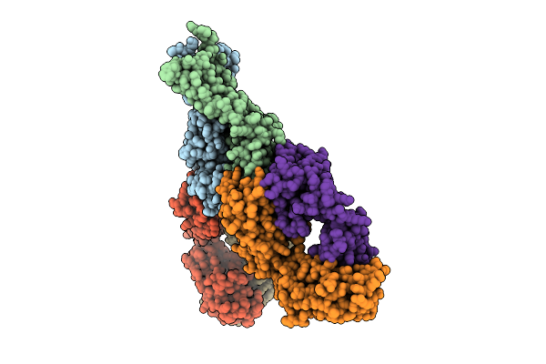
Cryoem Structure Of Respiratory Syncytial Virus Polymerase In Complex With Novel Non-Nucleoside Inhibitor Compound 16
Organism: Human respiratory syncytial virus, Synthetic construct
Method: ELECTRON MICROSCOPY Release Date: 2025-09-24 Classification: VIRAL PROTEIN,TRANSFERASE/INHIBITOR |
 |
Cryoem Map Of Respiratory Syncytial Virus Polymerase With Non-Nucleoside Inhibitor Compound 21
Organism: Human respiratory syncytial virus, Synthetic construct
Method: ELECTRON MICROSCOPY Release Date: 2025-09-24 Classification: VIRAL PROTEIN/INHIBITOR |
 |
Organism: Escherichia coli
Method: ELECTRON MICROSCOPY Release Date: 2025-09-17 Classification: RNA BINDING PROTEIN/DNA/RNA |
 |
Organism: Homo sapiens
Method: ELECTRON MICROSCOPY Release Date: 2025-09-03 Classification: LIGASE Ligands: ZN |
 |
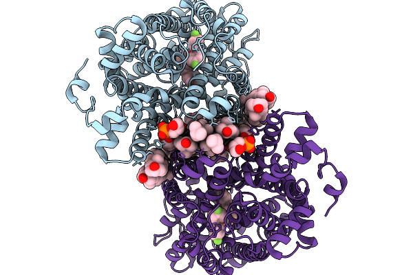
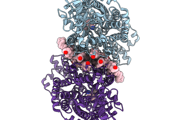
Cryo-Em Structure Of Neddylated Cul2-Rbx1-Elobc-Fem1B Homodimer Complexed With Fnip1 Degron
Organism: Homo sapiens, Escherichia coli k-12
Method: ELECTRON MICROSCOPY Release Date: 2025-09-03 Classification: LIGASE Ligands: ZN |
 |
Organism: Homo sapiens, Synthetic construct
Method: ELECTRON MICROSCOPY Release Date: 2025-08-13 Classification: STRUCTURAL PROTEIN/DNA |