
Search Count: 253
 |
Crystal Structure Of Human Prkcbp1 Zinc Finger Mynd-Type Containing 8 With Pentaglutamate Tag
Organism: Homo sapiens
Method: X-RAY DIFFRACTION Release Date: 2025-12-17 Classification: TRANSCRIPTION Ligands: EDO, ZN |
 |
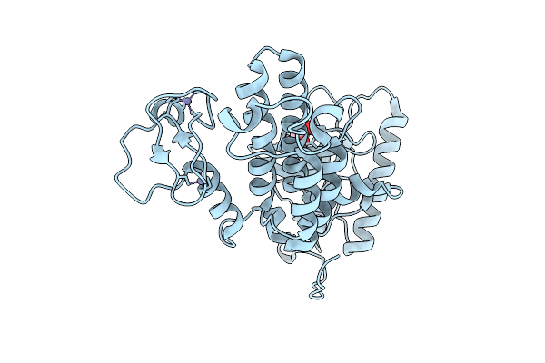
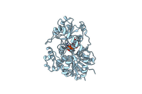
Organism: Pseudomonas aeruginosa
Method: X-RAY DIFFRACTION Release Date: 2025-12-10 Classification: SIGNALING PROTEIN Ligands: SO4 |
 |
Bromodomain Containing Protein 1 With Crystal Epitope Mutations P566E:V569R
Organism: Homo sapiens
Method: X-RAY DIFFRACTION Release Date: 2025-12-10 Classification: GENE REGULATION |
 |
Jumonji Domain-Containing Protein 2B With Crystallization Epitope Mutations L916G:R917A:A918D
Organism: Homo sapiens
Method: X-RAY DIFFRACTION Release Date: 2025-12-10 Classification: OXIDOREDUCTASE Ligands: EDO, SO4 |
 |
Protease From Norovirus Sydney Gii.4 Strain With Crystallization Epitope Mutation H50Y
Organism: Norovirus sydney 2212
Method: X-RAY DIFFRACTION Release Date: 2025-12-10 Classification: VIRAL PROTEIN |
 |
Jumonji Domain-Containing Protein 2B With Crystallization Epitope Mutations L916G:R917A:A918D
Organism: Homo sapiens
Method: X-RAY DIFFRACTION Release Date: 2025-12-03 Classification: OXIDOREDUCTASE Ligands: EDO, PEG, NH4 |
 |
Jumonji Domain-Containing Protein 2D With Crystallization Epitope Mutation Q41R
Organism: Homo sapiens
Method: X-RAY DIFFRACTION Release Date: 2025-11-26 Classification: OXIDOREDUCTASE Ligands: ZN, NI |
 |
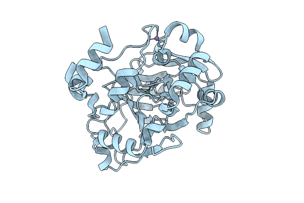
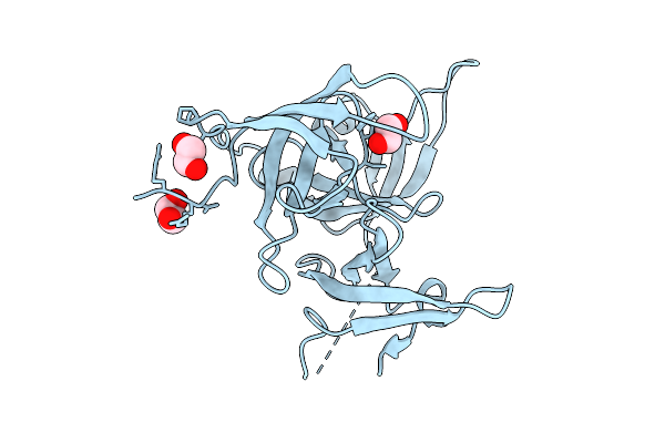
Human Prkcbp1 Zinc Finger Mynd-Type Containing 8 With Crystallization Epitope Mutations N221R:M226H
Organism: Homo sapiens
Method: X-RAY DIFFRACTION Release Date: 2025-11-26 Classification: TRANSFERASE Ligands: ZN |
 |
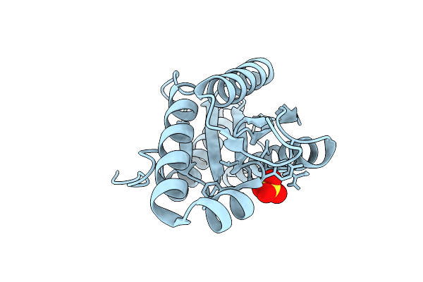
Organism: Homo sapiens
Method: X-RAY DIFFRACTION Release Date: 2025-11-26 Classification: CELL CYCLE Ligands: EDO |
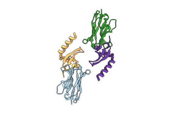 |
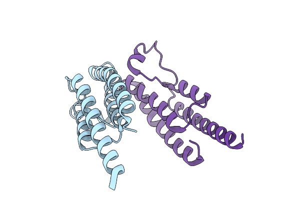
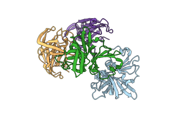
Organism: Lama glama, Homo sapiens
Method: X-RAY DIFFRACTION Release Date: 2025-11-05 Classification: GENE REGULATION |
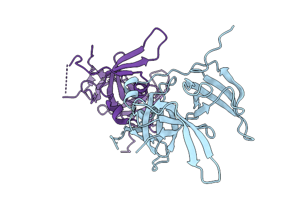 |
Spindlin Family Member 4 With Crystallization Epitope Mutations G129S:H131E
Organism: Homo sapiens
Method: X-RAY DIFFRACTION Release Date: 2025-11-05 Classification: PEPTIDE BINDING PROTEIN |
 |
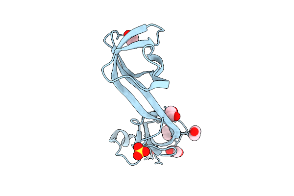
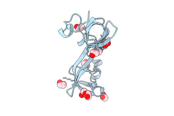
Organism: Homo sapiens
Method: X-RAY DIFFRACTION Release Date: 2025-10-29 Classification: HYDROLASE Ligands: CA, A1BM0 |
 |
Organism: Homo sapiens
Method: X-RAY DIFFRACTION Release Date: 2025-10-29 Classification: HYDROLASE Ligands: CA, PGE, A1BMX |
 |
Organism: Homo sapiens
Method: X-RAY DIFFRACTION Release Date: 2025-10-29 Classification: HYDROLASE Ligands: CA, PGE, EDO, A1BMY |
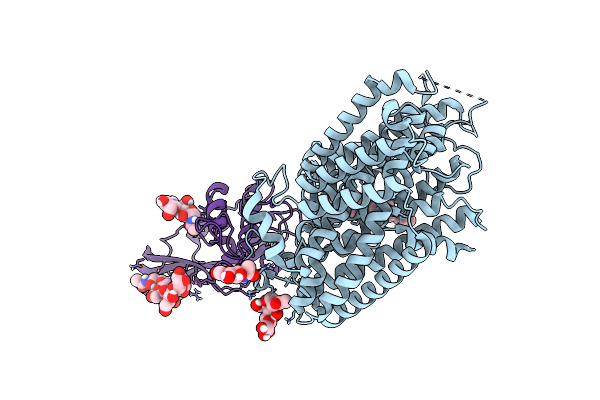 |
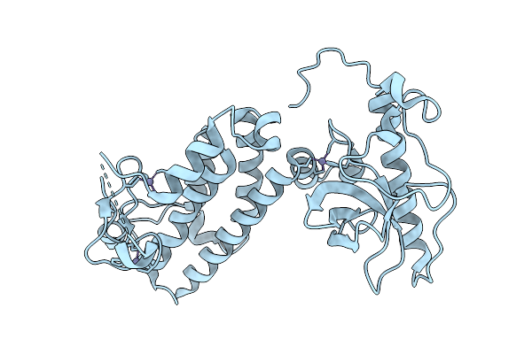
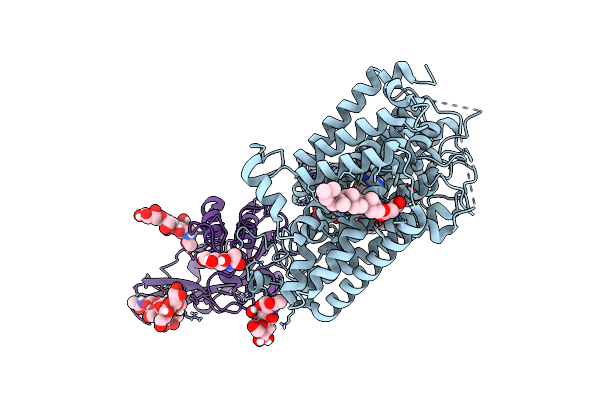
Cryo-Em Structure Of Mmcat1 Bound With Frmlv-Rbd In The Apo Inward-Open State
Organism: Mus musculus, Murine leukemia virus
Method: ELECTRON MICROSCOPY Release Date: 2025-07-02 Classification: MEMBRANE PROTEIN Ligands: NAG, Y01 |
 |
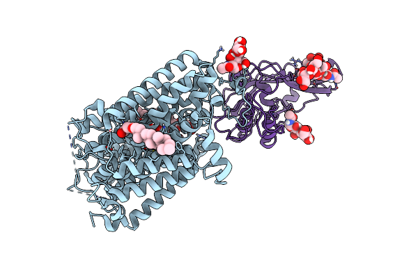
Cryo-Em Structure Of Mmcat1 Bound With Frmlv-Rbd In The Arginine-Bound Inward-Occluded State
Organism: Mus musculus, Murine leukemia virus
Method: ELECTRON MICROSCOPY Release Date: 2025-07-02 Classification: MEMBRANE PROTEIN Ligands: NAG, Y01, ARG |
 |
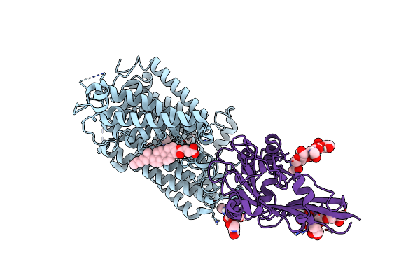
Cryo-Em Structure Of Mmcat1 Bound With Frmlv-Rbd In The Lysine-Bound Inward-Occluded State
Organism: Mus musculus, Murine leukemia virus
Method: ELECTRON MICROSCOPY Release Date: 2025-07-02 Classification: MEMBRANE PROTEIN Ligands: LYS, Y01 |
 |
Cryo-Em Structure Of Mmcat1 Bound With Frmlv-Rbd In The Ornithine-Bound Inward-Occluded State
Organism: Mus musculus, Murine leukemia virus
Method: ELECTRON MICROSCOPY Release Date: 2025-07-02 Classification: MEMBRANE PROTEIN Ligands: Y01, ORN |
 |
Organism: Homo sapiens
Method: X-RAY DIFFRACTION Release Date: 2025-06-25 Classification: CELL CYCLE Ligands: A1A6M, CL, MG |
 |
Organism: Cicer arietinum
Method: X-RAY DIFFRACTION Release Date: 2025-06-25 Classification: PLANT PROTEIN Ligands: UDP |