
Search Count: 22
 |
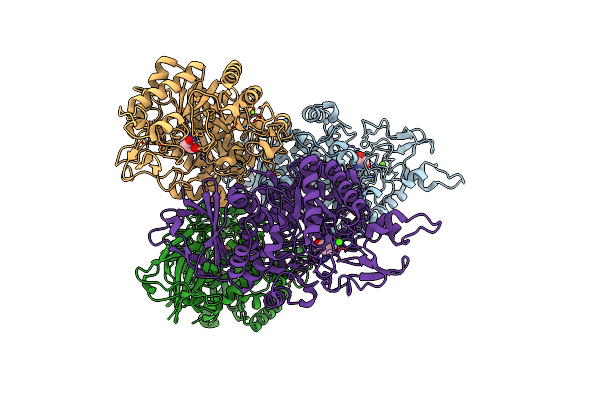
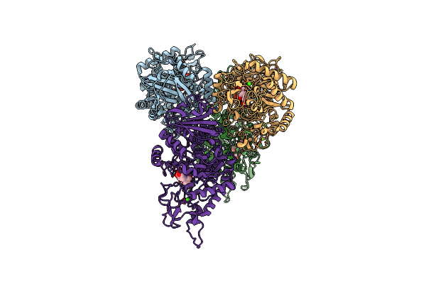
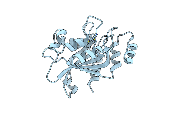
Human Topoisomerase 2 Alpha Atpase Domain Bound To Non-Hydrolyzable Atp Analog Amppnp
Organism: Homo sapiens
Method: X-RAY DIFFRACTION Release Date: 2025-06-25 Classification: ISOMERASE Ligands: ANP, MG |
 |
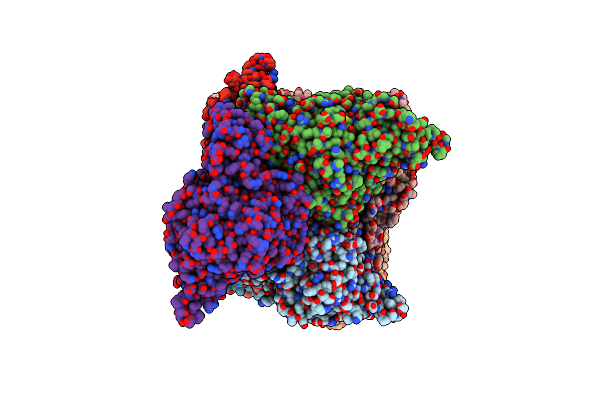
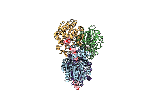
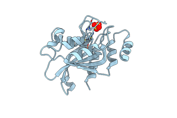
Human Topoisomerase 2 Alpha Atpase Domain Bound To Bns22 And Non-Hydrolyzable Atp Analog Amppnp
Organism: Homo sapiens
Method: X-RAY DIFFRACTION Release Date: 2025-06-25 Classification: ISOMERASE/INHIBITOR Ligands: ANP, MG, A1ASD, CL, GOL |
 |
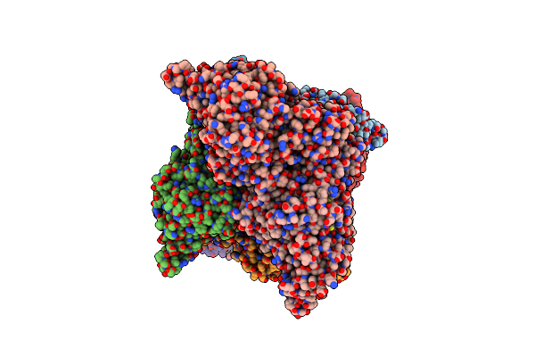
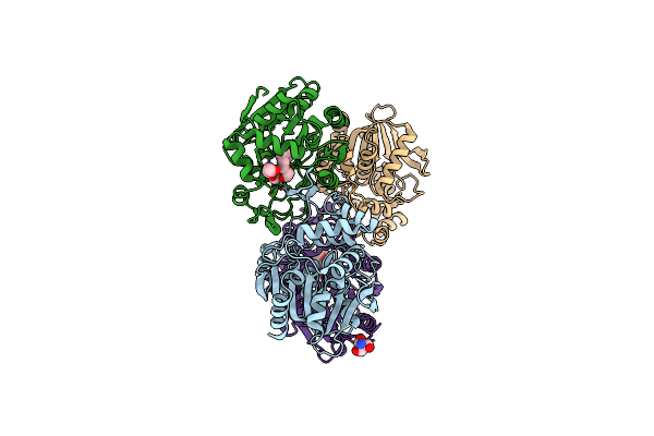
Human Topoisomerase 2 Beta Atpase Domain Bound To Non-Hydrolyzable Atp Analog Amppnp
Organism: Homo sapiens
Method: X-RAY DIFFRACTION Release Date: 2025-06-25 Classification: ISOMERASE Ligands: ANP, MG |
 |
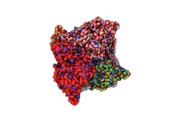
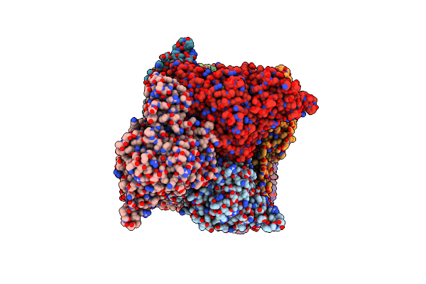
Human Topoisomerase 2 Alpha Atpase Domain Bound To Obex 5C And Non-Hydrolyzable Atp Analog Amppnp
Organism: Homo sapiens
Method: X-RAY DIFFRACTION Release Date: 2025-06-25 Classification: ISOMERASE/INHIBITOR Ligands: ANP, MG, A1ASE, GOL, CL |
 |
Human Topoisomerase 2 Beta Atpase Domain Bound To Bns22 And Non-Hydrolyzable Atp Analog Amppnp
Organism: Homo sapiens
Method: X-RAY DIFFRACTION Release Date: 2025-06-25 Classification: ISOMERASE/INHIBITOR Ligands: ANP, A1ASD, GOL, MG |
 |
Human Topoisomerase 2 Alpha Atpase Domain Bound To Topobexin And Non-Hydrolyzable Atp Analog Amppnp
Organism: Homo sapiens
Method: X-RAY DIFFRACTION Release Date: 2025-06-25 Classification: ISOMERASE/INHIBITOR Ligands: ANP, MG, A1ASC, CL, GOL |
 |
Human Topoisomerase 2 Beta Atpase Domain Bound To Obex 5C And Non-Hydrolyzable Atp Analog Amppnp
Organism: Homo sapiens
Method: X-RAY DIFFRACTION Release Date: 2025-06-25 Classification: ISOMERASE/INHIBITOR Ligands: ANP, A1ASE, MG |
 |
Human Topoisomerase 2 Beta Atpase Domain Bound To Topobexin And Non-Hydrolyzable Atp Analog Amppnp
Organism: Homo sapiens
Method: X-RAY DIFFRACTION Release Date: 2025-06-25 Classification: ISOMERASE/INHIBITOR Ligands: ANP, A1ASC, MG |
 |
Crystal Structure Of Trehalose Synthase Mutant N253C From Deinococcus Radiodurans
Organism: Deinococcus radiodurans
Method: X-RAY DIFFRACTION Resolution:2.65 Å Release Date: 2025-03-19 Classification: ISOMERASE Ligands: CA, MG, TRS |
 |
Crystal Structure Of Trehalose Synthase Mutant N253E From Deinococcus Radiodurans
Organism: Deinococcus radiodurans (strain atcc 13939 / dsm 20539 / jcm 16871 / ccug 27074 / lmg 4051 / nbrc 15346 / ncimb 9279 / vkm b-1422 / r1)
Method: X-RAY DIFFRACTION Resolution:3.04 Å Release Date: 2025-01-01 Classification: ISOMERASE Ligands: CA, MG, TRS |
 |
Crystal Structure Of Trehalose Synthase Mutant N253Q From Deinococcus Radiodurans
Organism: Deinococcus radiodurans (strain atcc 13939 / dsm 20539 / jcm 16871 / ccug 27074 / lmg 4051 / nbrc 15346 / ncimb 9279 / vkm b-1422 / r1)
Method: X-RAY DIFFRACTION Resolution:2.99 Å Release Date: 2025-01-01 Classification: ISOMERASE Ligands: CA, MG, TRS |
 |
Crystal Structure Of Trehalose Synthase Mutant N253T From Deinococcus Radiodurans
Organism: Deinococcus radiodurans (strain atcc 13939 / dsm 20539 / jcm 16871 / ccug 27074 / lmg 4051 / nbrc 15346 / ncimb 9279 / vkm b-1422 / r1)
Method: X-RAY DIFFRACTION Resolution:2.53 Å Release Date: 2025-01-01 Classification: ISOMERASE Ligands: CA, MG, TRS |
 |
Crystal Structure Of Trehalose Synthase Mutant R148A From Deinococcus Radiodurans
Organism: Deinococcus radiodurans
Method: X-RAY DIFFRACTION Resolution:2.32 Å Release Date: 2025-01-01 Classification: ISOMERASE Ligands: CA, MG, TRS |
 |
Crystal Structure Of Trehalose Synthase From Deinococcus Radiodurans Complexed With Validoxylamine A (Vaa)
Organism: Deinococcus radiodurans r1 = atcc 13939 = dsm 20539
Method: X-RAY DIFFRACTION Resolution:2.83 Å Release Date: 2025-01-01 Classification: ISOMERASE Ligands: CA, MG, VDM |
 |
Crystal Structure Of Trehalose Synthase Mutamt E324D From Deinococcus Radiodurans Complexed With Validoxylamine A (Vaa)
Organism: Deinococcus radiodurans (strain atcc 13939 / dsm 20539 / jcm 16871 / ccug 27074 / lmg 4051 / nbrc 15346 / ncimb 9279 / vkm b-1422 / r1)
Method: X-RAY DIFFRACTION Resolution:2.90 Å Release Date: 2025-01-01 Classification: ISOMERASE Ligands: CA, MG, VDM |
 |
Organism: Bacillus thuringiensis
Method: X-RAY DIFFRACTION Resolution:1.42 Å Release Date: 2024-12-11 Classification: HYDROLASE Ligands: P33, BTB |
 |
S102A Mutant Of Poly(3-Hydroxybutyrate) Depolymerase Phaz From Bacillus Thuringiensis
Organism: Bacillus thuringiensis
Method: X-RAY DIFFRACTION Resolution:1.90 Å Release Date: 2024-12-11 Classification: HYDROLASE Ligands: P33, TRS |
 |
Crystal Structure Of Trehalose Synthase Mutant N253H From Deinococcus Radiodurans
Organism: Deinococcus radiodurans
Method: X-RAY DIFFRACTION Resolution:2.97 Å Release Date: 2024-09-04 Classification: ISOMERASE Ligands: CA, MG, TRS |
 |
Organism: Aspergillus oryzae rib40
Method: X-RAY DIFFRACTION Resolution:1.44 Å Release Date: 2021-07-14 Classification: BIOSYNTHETIC PROTEIN Ligands: ZN, SO4 |
 |
Crystal Structure Of Aspergillus Oryzae Rib2 Deaminase (C-Terminal Deletion Mutant) At Ph 4.6
Organism: Aspergillus oryzae rib40
Method: X-RAY DIFFRACTION Resolution:1.70 Å Release Date: 2021-07-14 Classification: BIOSYNTHETIC PROTEIN Ligands: ZN |