
Search Count: 18
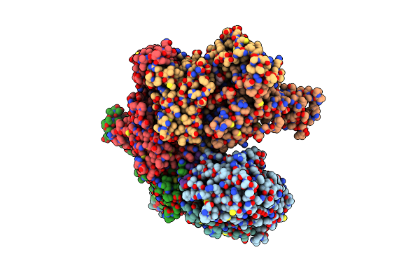 |
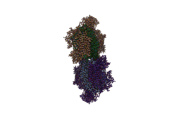
Organism: Homo sapiens
Method: ELECTRON MICROSCOPY Release Date: 2024-09-04 Classification: HYDROLASE Ligands: MG, ATP, ADP |
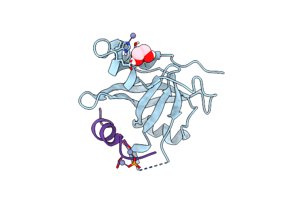 |
Organism: Saccharomyces cerevisiae
Method: X-RAY DIFFRACTION Resolution:1.58 Å Release Date: 2023-09-27 Classification: PROTEIN BINDING Ligands: EDO, ZN |
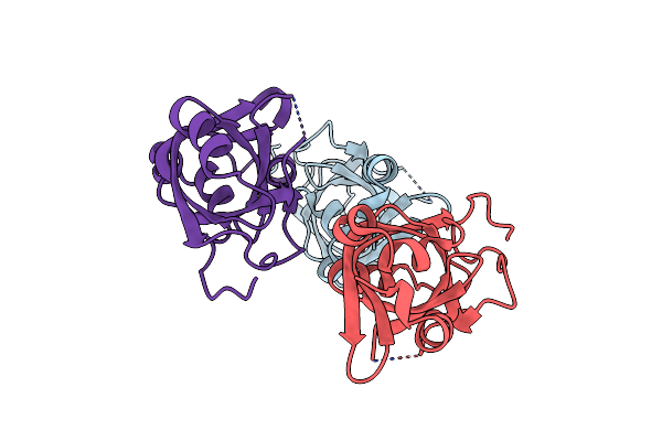 |
Organism: Saccharomyces cerevisiae
Method: X-RAY DIFFRACTION Resolution:1.55 Å Release Date: 2023-09-27 Classification: PROTEIN BINDING |
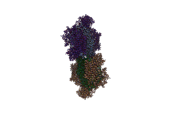 |
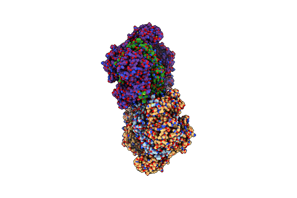
Organism: Saccharomyces cerevisiae s288c
Method: ELECTRON MICROSCOPY Release Date: 2020-11-11 Classification: HYDROLASE Ligands: ANP, ZN, MG |
 |
Organism: Saccharomyces cerevisiae s288c
Method: ELECTRON MICROSCOPY Release Date: 2020-11-11 Classification: HYDROLASE Ligands: ANP, ZN, MG |
 |
Organism: Saccharomyces cerevisiae s288c
Method: ELECTRON MICROSCOPY Release Date: 2020-11-11 Classification: HYDROLASE Ligands: ANP, ZN |
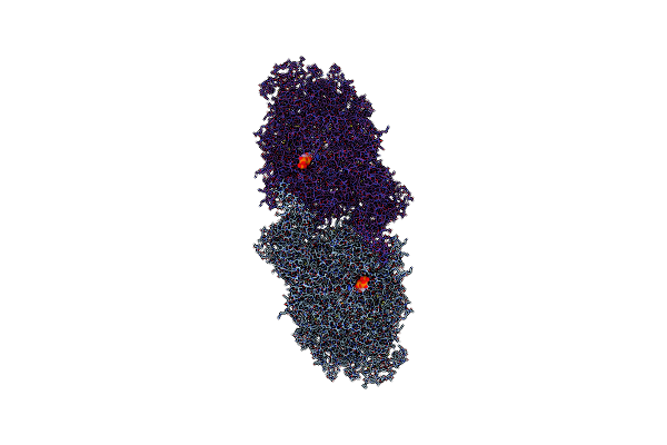 |
Organism: Saccharomyces cerevisiae
Method: ELECTRON MICROSCOPY Release Date: 2019-10-30 Classification: HYDROLASE Ligands: ANP, MG |
 |
Organism: Saccharomyces cerevisiae (strain atcc 204508 / s288c), Synthetic construct
Method: ELECTRON MICROSCOPY Release Date: 2018-12-19 Classification: DNA BINDING PROTEIN |
 |
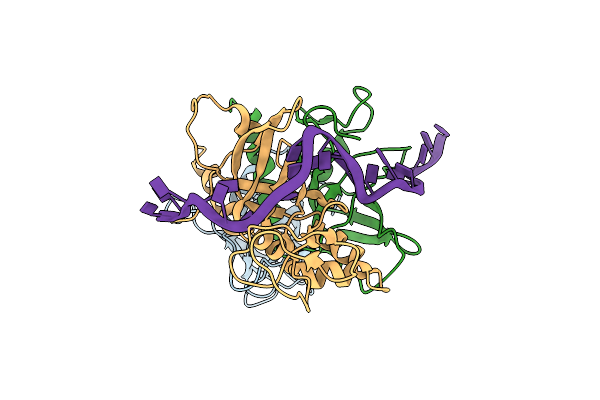
Organism: Mus musculus
Method: X-RAY DIFFRACTION Resolution:2.23 Å Release Date: 2017-01-11 Classification: SIGNALING PROTEIN |
 |
Structural Plasticity Of Cid1 Provides A Basis For Its Rna Terminal Uridylyl Transferase Activity
Organism: Schizosaccharomyces pombe
Method: X-RAY DIFFRACTION Resolution:1.74 Å Release Date: 2015-03-18 Classification: TRANSFERASE Ligands: GOL |
 |
Structural Plasticity Of Cid1 Provides A Basis For Its Rna Terminal Uridylyl Transferase Activity
Organism: Schizosaccharomyces pombe
Method: X-RAY DIFFRACTION Resolution:2.52 Å Release Date: 2015-03-18 Classification: TRANSFERASE |
 |
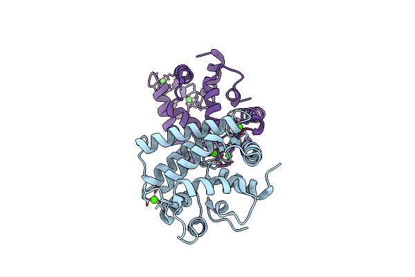
X-Ray Structure Of A Mutant (T61D) Of Calexcitin - A Neuronal Calcium-Signalling Protein
Organism: Doryteuthis pealeii
Method: X-RAY DIFFRACTION Resolution:2.00 Å Release Date: 2014-10-29 Classification: SIGNALING PROTEIN Ligands: CA |
 |
X-Ray Structure Of A Mutant (T188D) Of Calexcitin - A Neuronal Calcium-Signalling Protein
Organism: Doryteuthis pealeii
Method: X-RAY DIFFRACTION Resolution:2.00 Å Release Date: 2014-10-29 Classification: SIGNALING PROTEIN Ligands: CA |
 |
X-Ray Structure Of A Double Mutant Of Calexcitin - A Neuronal Calcium-Signalling Protein
Organism: Doryteuthis pealeii
Method: X-RAY DIFFRACTION Resolution:2.90 Å Release Date: 2014-10-29 Classification: SIGNALING PROTEIN Ligands: CA |
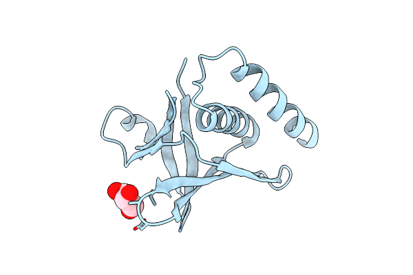 |
Structural And Functional Characterisation Of The Kindlin-1 Pleckstrin Homology Domain
Organism: Mus musculus
Method: X-RAY DIFFRACTION Resolution:2.10 Å Release Date: 2012-11-14 Classification: CELL ADHESION Ligands: GOL |
 |
Structural Basis For The Activity Of A Cytoplasmic Rna Terminal U-Transferase
Organism: Schizosaccharomyces pombe 972h-
Method: X-RAY DIFFRACTION Resolution:3.20 Å Release Date: 2012-07-04 Classification: TRANSFERASE Ligands: ACT |
 |
Structural Basis For The Activity Of A Cytoplasmic Rna Terminal U-Transferase
Organism: Schizosaccharomyces pombe 972h-
Method: X-RAY DIFFRACTION Resolution:3.02 Å Release Date: 2012-07-04 Classification: TRANSFERASE Ligands: UTP |
 |
Structural Basis For The Activity Of A Cytoplasmic Rna Terminal U-Transferase
Organism: Schizosaccharomyces pombe 972h-
Method: X-RAY DIFFRACTION Resolution:2.60 Å Release Date: 2012-07-04 Classification: TRANSFERASE Ligands: ACT, GOL |