
Search Count: 23
 |
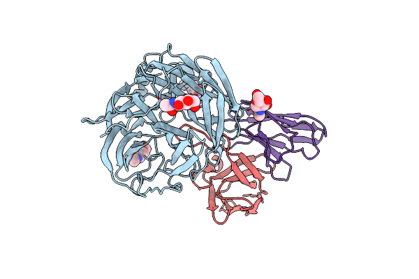
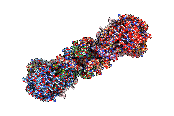
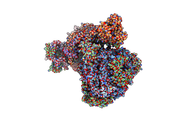
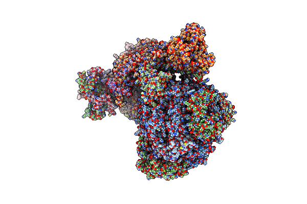
Ncs.1 Fab In Complex With N5 Na Of A/Shorebird/Delaware Bay/309/2016 (Db16, H10N5) -- 4 Fabs
Organism: Homo sapiens, Influenza a virus
Method: ELECTRON MICROSCOPY Release Date: 2025-08-13 Classification: VIRAL PROTEIN Ligands: NAG, CA |
 |
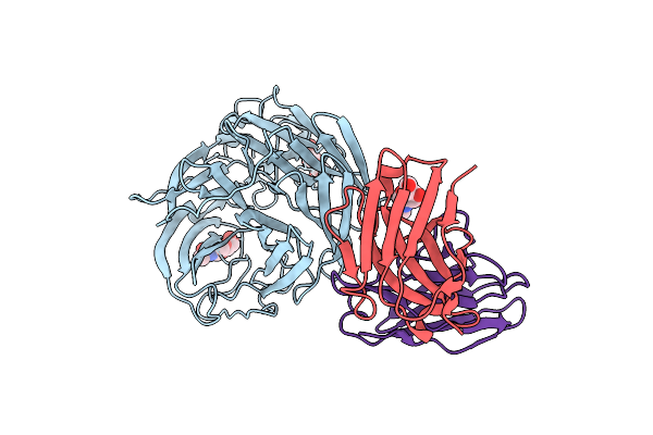
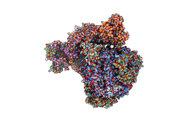
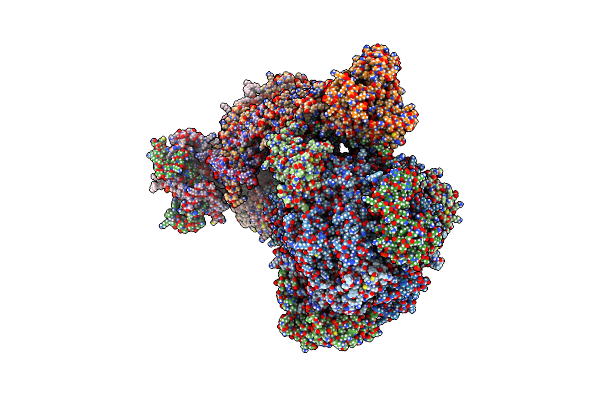
Ncs.1 Fab In Complex With N5 Na Of A/Shorebird/Delaware Bay/309/2016 (Db16, H10N5) -- 3 Fabs
Organism: Influenza a virus, Homo sapiens
Method: ELECTRON MICROSCOPY Release Date: 2025-08-13 Classification: VIRAL PROTEIN Ligands: NAG, CA |
 |
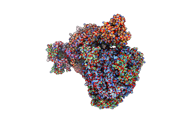
Ncs.1.1 Fab In Complex With The Snap Of A/California/04/2009 (Ca09, H1N1) -- 4 Fabs [C4 Reconstruction]
Organism: Influenza a virus, Homo sapiens
Method: ELECTRON MICROSCOPY Release Date: 2025-08-13 Classification: VIRAL PROTEIN Ligands: NAG, CA |
 |
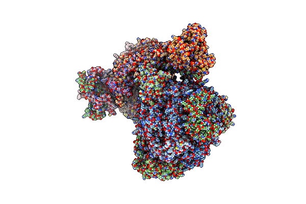
Ncs.1.1 Fab In Complex With The Snap Of A/California/04/2009 (Ca09, H1N1) -- 4 Fabs [C1 Reconstruction]
Organism: Influenza a virus, Homo sapiens
Method: ELECTRON MICROSCOPY Release Date: 2025-08-13 Classification: VIRAL PROTEIN Ligands: NAG, CA |
 |
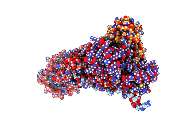
Structure Of Human Nds.1 Fab And 1G01 Fab In Complex With Influenza Virus Neuraminidase From A/Indiana/10/2011 (H3N2V), Based On Consensus Cryo-Em Map With Only Fab 1G01 Resolved
Organism: Homo sapiens, Influenza a virus (a/indiana/10/2011(h3n2))
Method: ELECTRON MICROSCOPY Release Date: 2024-02-28 Classification: VIRAL PROTEIN/IMMUNE SYSTEM Ligands: NAG |
 |
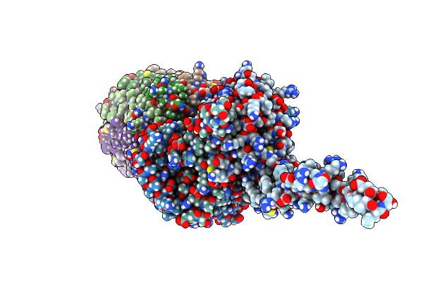
Structure Of Human Nds.1 Fab And 1G01 Fab In Complex With Influenza Virus Neuraminidase From A/Indiana/10/2011 (H3N2V)
Organism: Homo sapiens, Influenza a virus (a/indiana/10/2011(h3n2))
Method: ELECTRON MICROSCOPY Release Date: 2024-02-28 Classification: VIRAL PROTEIN/IMMUNE SYSTEM Ligands: NAG |
 |
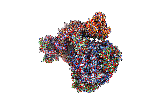
Structure Of Human Nds.3 Fab In Complex With Influenza Virus Neuraminidase From A/Darwin/09/2021 (H3N2)
Organism: Influenza a virus, Homo sapiens
Method: ELECTRON MICROSCOPY Release Date: 2024-02-28 Classification: VIRAL PROTEIN/IMMUNE SYSTEM Ligands: NAG |
 |
Organism: Trypanosoma brucei brucei
Method: ELECTRON MICROSCOPY Release Date: 2022-12-07 Classification: MEMBRANE PROTEIN Ligands: ATP, MG, CDL, ADP, LMT, Q7G, PEE, PC1, UTP |
 |
Organism: Trypanosoma brucei brucei
Method: ELECTRON MICROSCOPY Release Date: 2022-10-26 Classification: MEMBRANE PROTEIN Ligands: CDL, LMT, Q7G, PEE, PC1 |
 |
Organism: Trypanosoma brucei brucei
Method: ELECTRON MICROSCOPY Release Date: 2022-10-26 Classification: MEMBRANE PROTEIN |
 |
Organism: Trypanosoma brucei brucei
Method: ELECTRON MICROSCOPY Release Date: 2022-10-26 Classification: MEMBRANE PROTEIN Ligands: UTP |
 |
Rotational State 1A Of The Trypanosoma Brucei Mitochondrial Atp Synthase Dimer
Organism: Trypanosoma brucei brucei
Method: ELECTRON MICROSCOPY Release Date: 2022-10-26 Classification: MEMBRANE PROTEIN Ligands: ATP, MG, ADP, UTP, CDL, PEE, LMT, Q7G, PC1 |
 |
Rotational State 1B Of The Trypanosoma Brucei Mitochondrial Atp Synthase Dimer
Organism: Trypanosoma brucei brucei
Method: ELECTRON MICROSCOPY Release Date: 2022-10-26 Classification: MEMBRANE PROTEIN Ligands: ATP, MG, ADP, UTP, CDL, 3PE, PC1, LMT, Q7G |
 |
Rotational State 1C Of The Trypanosoma Brucei Mitochondrial Atp Synthase Dimer
Organism: Trypanosoma brucei brucei
Method: ELECTRON MICROSCOPY Release Date: 2022-10-26 Classification: MEMBRANE PROTEIN Ligands: ATP, MG, ADP, UTP, CDL, 3PE, LMT, Q7G, PC1 |
 |
Rotational State 1D Of The Trypanosoma Brucei Mitochondrial Atp Synthase Dimer
Organism: Trypanosoma brucei brucei
Method: ELECTRON MICROSCOPY Release Date: 2022-10-26 Classification: MEMBRANE PROTEIN Ligands: CDL, PEE, PC1, LMT, Q7G, ATP, MG, ADP, UTP |
 |
Rotational State 1E Of The Trypanosoma Brucei Mitochondrial Atp Synthase Dimer
Organism: Trypanosoma brucei brucei
Method: ELECTRON MICROSCOPY Release Date: 2022-10-26 Classification: MEMBRANE PROTEIN Ligands: ATP, MG, ADP, UTP, CDL, PEE, PC1, LMT, Q7G |
 |
Rotational State 2A Of The Trypanosoma Brucei Mitochondrial Atp Synthase Dimer
Organism: Trypanosoma brucei brucei
Method: ELECTRON MICROSCOPY Release Date: 2022-10-26 Classification: MEMBRANE PROTEIN Ligands: CDL, PEE, LMT, Q7G, PC1, UTP, ATP, MG, ADP |
 |
Rotational State 2B Of The Trypanosoma Brucei Mitochondrial Atp Synthase Dimer
Organism: Trypanosoma brucei brucei
Method: ELECTRON MICROSCOPY Release Date: 2022-10-26 Classification: MEMBRANE PROTEIN Ligands: CDL, PEE, LMT, Q7G, PC1, UTP, ATP, MG, ADP |
 |
Rotational State 2C Of The Trypanosoma Brucei Mitochondrial Atp Synthase Dimer
Organism: Trypanosoma brucei brucei
Method: ELECTRON MICROSCOPY Release Date: 2022-10-26 Classification: MEMBRANE PROTEIN Ligands: CDL, PEE, LMT, Q7G, PC1, UTP, ATP, MG, ADP |
 |
Organism: Trypanosoma brucei brucei
Method: ELECTRON MICROSCOPY Release Date: 2022-10-26 Classification: MEMBRANE PROTEIN Ligands: CDL, PEE, LMT, Q7G, PC1, UTP, ATP, MG, ADP |