
Search Count: 16
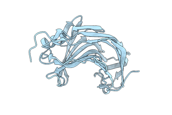 |
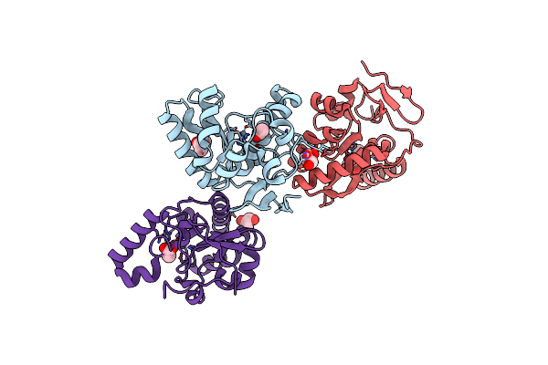
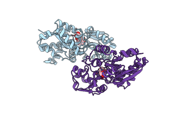
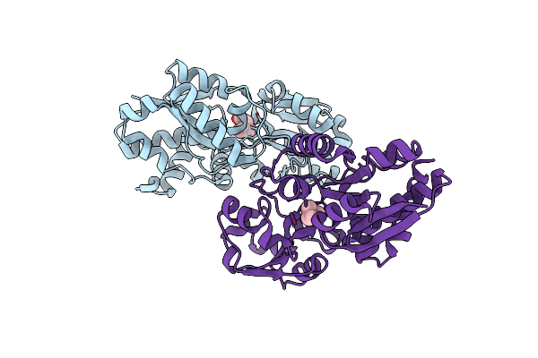
Crystal Structure Of The Pathogen-Secreted Apoplastic Gh12 Xyloglucan-Specific Endoglucanase Xeg1
Organism: Phytophthora sojae (strain p6497)
Method: X-RAY DIFFRACTION Release Date: 2025-12-10 Classification: HYDROLASE |
 |
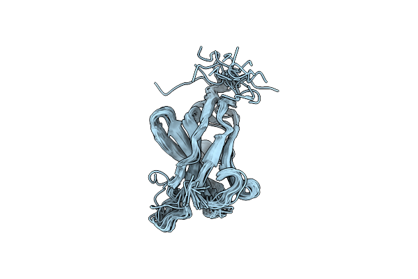
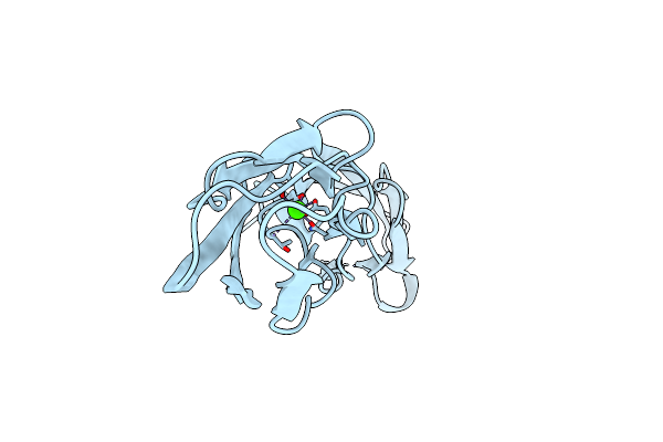
Solution Nmr Structure Of Zinc Fingers 1-2 (Fragment 257-320) From Human Insulinoma-Associated Protein 1(Insm1)
Organism: Homo sapiens
Method: SOLUTION NMR Release Date: 2024-06-19 Classification: DNA BINDING PROTEIN Ligands: ZN |
 |
Crystal Structure Of Odorant Binding Protein 4 In The Natural Predator Chrysopa Pallens
Organism: Chrysopa pallens
Method: X-RAY DIFFRACTION Resolution:2.10 Å Release Date: 2019-10-16 Classification: STRUCTURAL PROTEIN |
 |
Organism: Vicugna pacos
Method: SOLUTION NMR Release Date: 2019-06-05 Classification: AFB1-BINDING PROTEIN |
 |
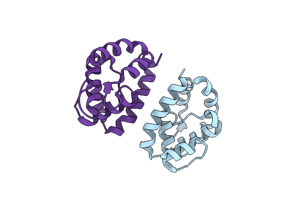
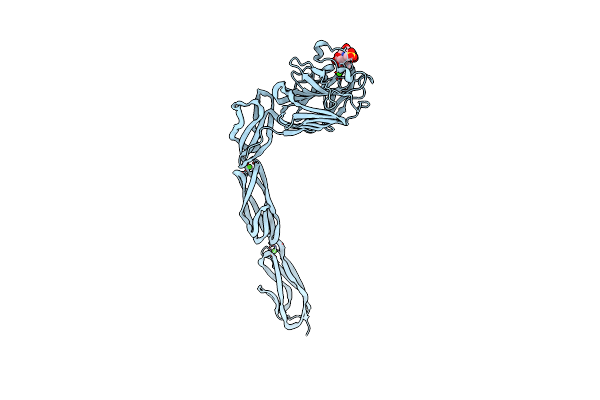
Crystal Structure Of An L,D-Carboxypeptidase Dacb From Streptococcus Pneumonia
Organism: Streptococcus pneumoniae
Method: X-RAY DIFFRACTION Resolution:1.71 Å Release Date: 2014-11-12 Classification: HYDROLASE Ligands: ZN, ACT, GOL |
 |
Organism: Escherichia coli
Method: SOLUTION NMR Release Date: 2014-08-20 Classification: TRANSFERASE Ligands: PO3 |
 |
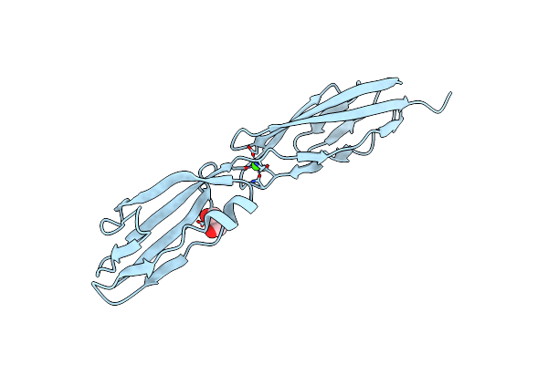
Crystal Structure Of The Ligand Binding Region Of Staphylococcal Adhesion Srap
Organism: Staphylococcus aureus
Method: X-RAY DIFFRACTION Resolution:2.05 Å Release Date: 2014-06-18 Classification: CELL ADHESION Ligands: CA, MES |
 |
Organism: Staphylococcus aureus
Method: X-RAY DIFFRACTION Resolution:2.10 Å Release Date: 2014-06-18 Classification: CELL ADHESION Ligands: CA, GOL |
 |
Organism: Staphylococcus aureus
Method: X-RAY DIFFRACTION Resolution:1.59 Å Release Date: 2014-06-18 Classification: Calcium Binding protein Ligands: CA, GOL |
 |
Organism: Staphylococcus aureus
Method: X-RAY DIFFRACTION Resolution:2.24 Å Release Date: 2014-06-18 Classification: Calcium Binding protein Ligands: CA |
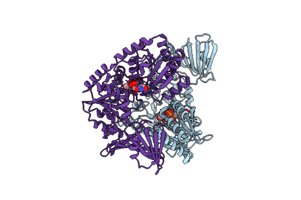 |
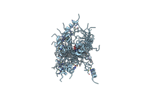
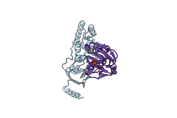
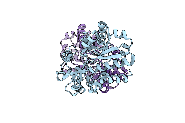
Crystal Structure Of The Pneumococcal O-Glcnac Transferase Gtfa In Complex With Udp And Glcnac
Organism: Streptococcus pneumoniae
Method: X-RAY DIFFRACTION Resolution:2.00 Å Release Date: 2014-06-18 Classification: TRANSFERASE Ligands: UDP, NAG |
 |
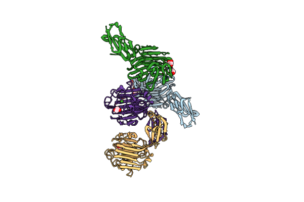
Structures Of The Substrate-Binding Protein Provide Insights Into The Multiple Compatible Solutes Binding Specificities Of Bacillus Subtilis Abc Transporter Opuc
Organism: Bacillus subtilis
Method: X-RAY DIFFRACTION Resolution:2.30 Å Release Date: 2011-05-11 Classification: TRANSPORT PROTEIN |
 |
Structures Of The Substrate-Binding Protein Provide Insights Into The Multiple Compatible Solutes Binding Specificities Of Bacillus Subtilis Abc Transporter Opuc
Organism: Bacillus subtilis
Method: X-RAY DIFFRACTION Resolution:2.70 Å Release Date: 2011-05-11 Classification: TRANSPORT PROTEIN Ligands: DCK |
 |
Structures Of The Substrate-Binding Protein Provide Insights Into The Multiple Compatible Solutes Binding Specificities Of Bacillus Subtilis Abc Transporter Opuc
Organism: Bacillus subtilis
Method: X-RAY DIFFRACTION Resolution:2.40 Å Release Date: 2011-05-11 Classification: TRANSPORT PROTEIN Ligands: BET |
 |
Structures Of The Substrate-Binding Protein Provide Insights Into The Multiple Compatible Solutes Binding Specificities Of Bacillus Subtilis Abc Transporter Opuc
Organism: Bacillus subtilis
Method: X-RAY DIFFRACTION Resolution:1.91 Å Release Date: 2011-05-11 Classification: TRANSPORT PROTEIN Ligands: CHT |
 |
Structures Of The Substrate-Binding Protein Provide Insights Into The Multiple Compatible Solutes Binding Specificities Of Bacillus Subtilis Abc Transporter Opuc
Organism: Bacillus subtilis
Method: X-RAY DIFFRACTION Resolution:2.10 Å Release Date: 2011-05-11 Classification: TRANSPORT PROTEIN Ligands: 4CS |