
Search Count: 4
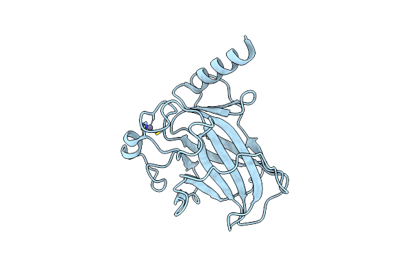 |
Organism: Homo sapiens
Method: X-RAY DIFFRACTION Resolution:1.30 Å Release Date: 2023-05-17 Classification: DNA BINDING PROTEIN Ligands: ZN |
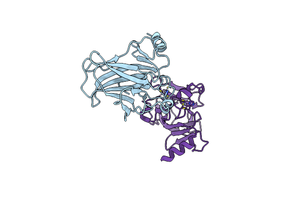 |
Organism: Homo sapiens
Method: X-RAY DIFFRACTION Resolution:2.50 Å Release Date: 2023-05-17 Classification: DNA BINDING PROTEIN Ligands: ZN |
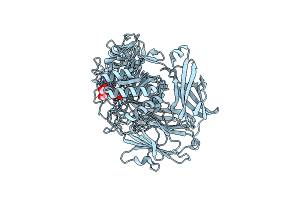 |
Crystal Structure Of Beta-Galactosidase Ii From Bacillus Circulans In Complex With Beta-D-Galactopyranosyl Disaccharide
Organism: Bacillus circulans
Method: X-RAY DIFFRACTION Resolution:2.00 Å Release Date: 2020-12-09 Classification: HYDROLASE Ligands: GLC, GAL |
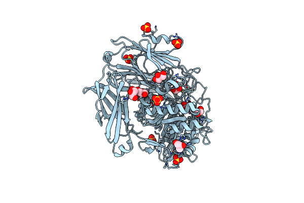 |
Organism: Bacillus circulans
Method: X-RAY DIFFRACTION Resolution:1.95 Å Release Date: 2020-12-09 Classification: HYDROLASE Ligands: GOL, SO4, CL |