
Search Count: 164
 |
Organism: Frankia casuarinae
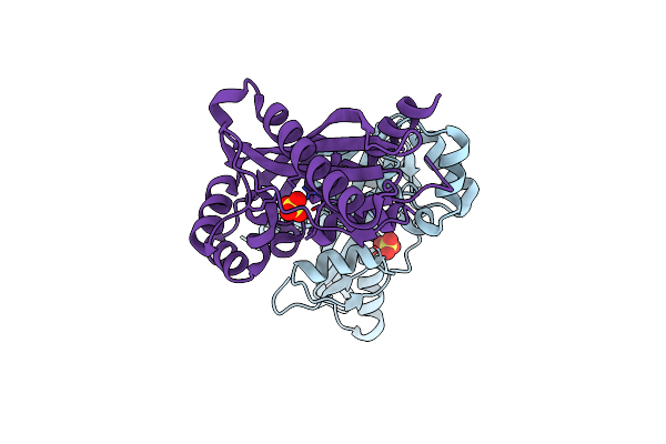
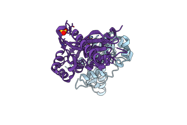
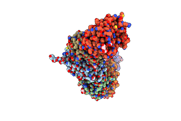
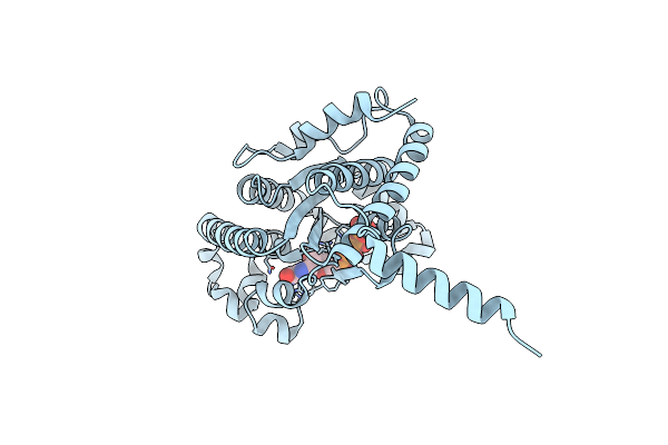
Method: X-RAY DIFFRACTION Release Date: 2025-01-15 Classification: OXIDOREDUCTASE Ligands: HEM, OXY |
 |
Organism: Pseudomonas rhizosphaerae
Method: X-RAY DIFFRACTION Release Date: 2025-01-15 Classification: OXIDOREDUCTASE Ligands: HEM, OXY, CL, SO4 |
 |
Organism: Pseudomonas fluorescens (strain atcc baa-477 / nrrl b-23932 / pf-5)
Method: X-RAY DIFFRACTION Release Date: 2025-01-15 Classification: OXIDOREDUCTASE Ligands: HEM, OXY |
 |
Crystal Structure Of Cib_12 Beta-Galactosidase From Cuniculiplasma Divulgatum
Organism: Cuniculiplasma divulgatum
Method: X-RAY DIFFRACTION Resolution:2.55 Å Release Date: 2024-07-24 Classification: HYDROLASE Ligands: GOL |
 |
Crystal Structure Of Cib_13 Beta-Galactosidase From Cuniculiplasma Divulgatum
Organism: Cuniculiplasma divulgatum
Method: X-RAY DIFFRACTION Resolution:2.22 Å Release Date: 2024-07-24 Classification: HYDROLASE Ligands: GOL, TRS |
 |
Organism: Clostridium carboxidivorans p7
Method: X-RAY DIFFRACTION Resolution:2.38 Å Release Date: 2024-03-13 Classification: TRANSPORT PROTEIN Ligands: CL |
 |
Crystal Structure Of Fluoroacetate Dehalogenase Daro3835 H274N Mutant With D107-Glycolyl Intermediate
Organism: Dechloromonas aromatica rcb
Method: X-RAY DIFFRACTION Resolution:2.00 Å Release Date: 2024-02-14 Classification: HYDROLASE |
 |
Organism: Dechloromonas aromatica rcb
Method: X-RAY DIFFRACTION Resolution:1.86 Å Release Date: 2023-09-06 Classification: HYDROLASE Ligands: CL |
 |
Crystal Structure Of A Gh12-2 Family Cellulase From Thermococcus Sp. 2319X1
Organism: Thermococcus sp. 2319x1
Method: X-RAY DIFFRACTION Resolution:2.55 Å Release Date: 2022-08-24 Classification: HYDROLASE Ligands: GOL, PE3, CA, CL |
 |
Organism: Dehalococcoidia bacterium
Method: X-RAY DIFFRACTION Resolution:2.92 Å Release Date: 2022-08-24 Classification: HYDROLASE |
 |
Crystal Structure Of Maf Domain Of Human N-Acetylserotonin O-Methyltransferase-Like Protein Soaked With Tfbq
Organism: Homo sapiens
Method: X-RAY DIFFRACTION Resolution:2.22 Å Release Date: 2021-06-23 Classification: CELL CYCLE Ligands: CL, SO4 |
 |
Crystal Structure Of Human N-Acetylserotonin O-Methyltransferase-Like Protein Soaked With Pdhptao
Organism: Homo sapiens
Method: X-RAY DIFFRACTION Resolution:2.61 Å Release Date: 2021-06-23 Classification: CELL CYCLE Ligands: SO4 |
 |
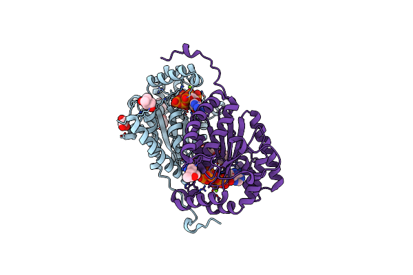
Crystal Structure Of The Adenylation (A) Domain Of The Carboxylate Reductase (Car) Gr01_22995 From Mycobacterium Chelonae
Organism: Mycobacterium chelonae
Method: X-RAY DIFFRACTION Resolution:1.97 Å Release Date: 2020-04-22 Classification: OXIDOREDUCTASE Ligands: SO4, CL, AMP, SIN, GOL |
 |
Crystal Structure Of Bh1352 2-Deoxyribose-5-Phosphate From Bacillus Halodurans, K184L Mutant
Organism: Bacillus halodurans c-125
Method: X-RAY DIFFRACTION Resolution:2.17 Å Release Date: 2019-10-23 Classification: LYASE Ligands: GOL |
 |
Crystal Structure Of Bh1352 2-Deoxyribose-5-Phosphate From Bacillus Halodurans
Organism: Bacillus halodurans (strain atcc baa-125 / dsm 18197 / ferm 7344 / jcm 9153 / c-125)
Method: X-RAY DIFFRACTION Resolution:2.50 Å Release Date: 2019-10-16 Classification: TRANSFERASE Ligands: GOL, TRS |
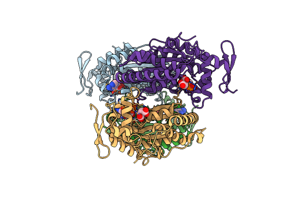 |
Crystal Structure Of Polyphosphate Kinase 2 Class I (Smc02148) In Complex With Adp
Organism: Rhizobium meliloti (strain 1021)
Method: X-RAY DIFFRACTION Resolution:1.87 Å Release Date: 2019-07-10 Classification: TRANSFERASE Ligands: ADP, MLT, AMP |
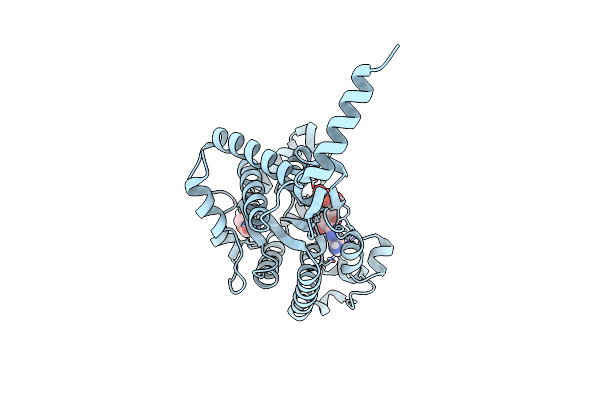 |
Crystal Structure Of Ppk2 Class Iii In Complex With Adp From Cytophaga Hutchinsonii Atcc 33406
Organism: Cytophaga hutchinsonii (strain atcc 33406 / ncimb 9469)
Method: X-RAY DIFFRACTION Resolution:1.89 Å Release Date: 2019-01-16 Classification: TRANSFERASE Ligands: ADP, GOL |
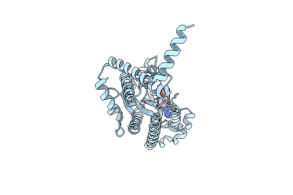 |
Crystal Structure Of Ppk2 Class Iii In The Complex With Amp From Cytophaga Hutchinsonii Atcc 33406
Organism: Cytophaga hutchinsonii (strain atcc 33406 / ncimb 9469)
Method: X-RAY DIFFRACTION Resolution:2.45 Å Release Date: 2019-01-16 Classification: TRANSFERASE Ligands: AMP, CL |
 |
Crystal Structure Of Ppk2 Class Iii In Complex With Guanosine 5-Tetraphosphate
Organism: Cytophaga hutchinsonii (strain atcc 33406 / ncimb 9469)
Method: X-RAY DIFFRACTION Resolution:2.65 Å Release Date: 2019-01-16 Classification: TRANSFERASE Ligands: BKP |
 |
Organism: Deinococcus radiodurans (strain atcc 13939 / dsm 20539 / jcm 16871 / lmg 4051 / nbrc 15346 / ncimb 9279 / r1 / vkm b-1422)
Method: X-RAY DIFFRACTION Resolution:1.81 Å Release Date: 2019-01-16 Classification: TRANSFERASE Ligands: ATP, MG, GOL, MPD, CL |