
Search Count: 16
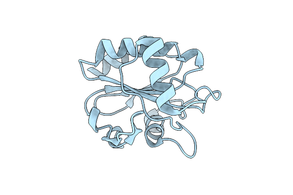 |
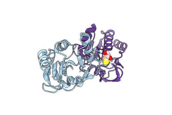
Crystal Structure Of C36S Mutant Glutathione Peroxidase Of Staphylococcus Aureus.
Organism: Staphylococcus aureus
Method: X-RAY DIFFRACTION Release Date: 2025-12-17 Classification: OXIDOREDUCTASE |
 |
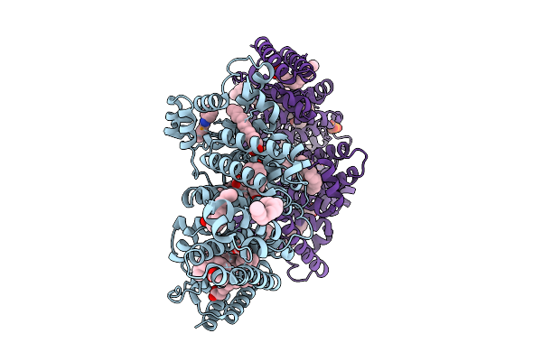
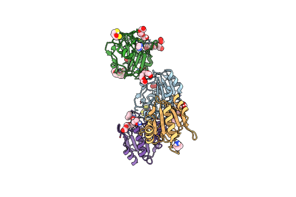
Crystal Structure Of Pseudomonas Aeruginosa Suhb Complexed With Gallic Acid In Monoclinic Space Group
Organism: Pseudomonas aeruginosa
Method: X-RAY DIFFRACTION Release Date: 2025-09-10 Classification: HYDROLASE Ligands: GOL, GDE, DTT |
 |
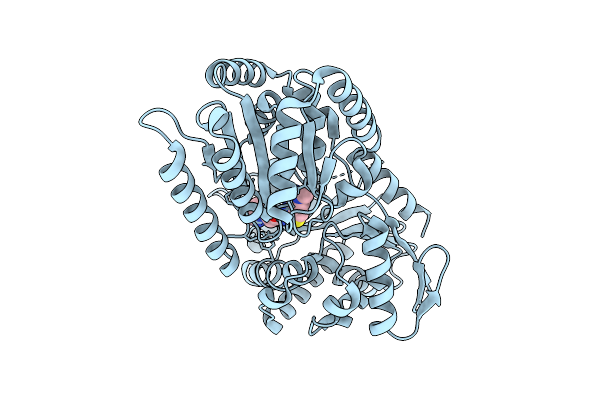
Organism: Homo sapiens
Method: X-RAY DIFFRACTION Release Date: 2025-09-10 Classification: TRANSPORT PROTEIN Ligands: 9JT, MYR, PO4 |
 |
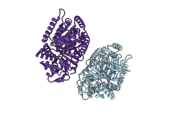
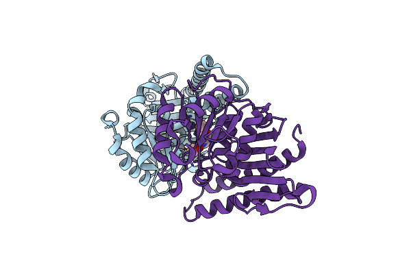
Crystal Structure Of Pseudomonas Aeruginosa Suhb Complexed With Gallic Acid In Orthorhombic Space Group
Organism: Pseudomonas aeruginosa pao1
Method: X-RAY DIFFRACTION Release Date: 2025-08-27 Classification: HYDROLASE Ligands: DTV, GDE |
 |
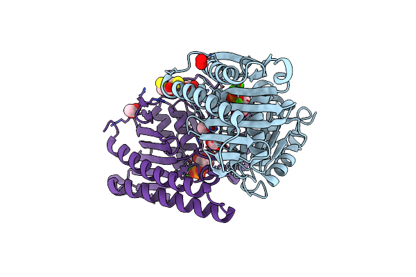
Structure Of Candida Albicans Trehalose-6-Phosphate Synthase In Complex With 4456
Organism: Candida albicans sc5314
Method: X-RAY DIFFRACTION Release Date: 2025-08-20 Classification: TRANSFERASE Ligands: A1BYI |
 |
Structure Of Candida Albicans Trehalose-6-Phosphate Synthase In Complex With Sj6675
Organism: Candida albicans sc5314
Method: X-RAY DIFFRACTION Release Date: 2025-08-20 Classification: TRANSFERASE Ligands: A1BYV |
 |
Crystal Structure Of Atypical Two Cysteine Thiol Peroxidase From Staphylococcus Aureus
Organism: Staphylococcus aureus
Method: X-RAY DIFFRACTION Release Date: 2025-06-18 Classification: OXIDOREDUCTASE Ligands: DTU |
 |
Crystal Structure Of N Terminal Deletion Mutant Of Staphylococcal Atypical Two-Cysteine Thiol Peroxidase Complexed With Substrate Analog
Organism: Staphylococcus aureus
Method: X-RAY DIFFRACTION Release Date: 2025-06-18 Classification: OXIDOREDUCTASE Ligands: PG4, PEO, A1L2G, IMD, DTO, DTU |
 |
Crystal Structure Of N-Terminal Deletion Mutant Of Staphylococcal Thiol Peroxidase
Organism: Staphylococcus aureus subsp. aureus nctc 8325
Method: X-RAY DIFFRACTION Resolution:1.80 Å Release Date: 2025-01-22 Classification: OXIDOREDUCTASE Ligands: DTU, PG4, GOL, SO4 |
 |
Organism: Pseudomonas aeruginosa pao1
Method: X-RAY DIFFRACTION Resolution:2.60 Å Release Date: 2024-10-02 Classification: HYDROLASE Ligands: ACT, IMD |
 |
Crystal Structure Of Pseudomonas Aeruginosa Suhb In Complex With D-Myo-Inositol-1-Phosphate
Organism: Pseudomonas aeruginosa pao1
Method: X-RAY DIFFRACTION Resolution:2.20 Å Release Date: 2024-09-18 Classification: HYDROLASE Ligands: IPD, ACT, PEO, GOL, DTU, CL, CA |
 |
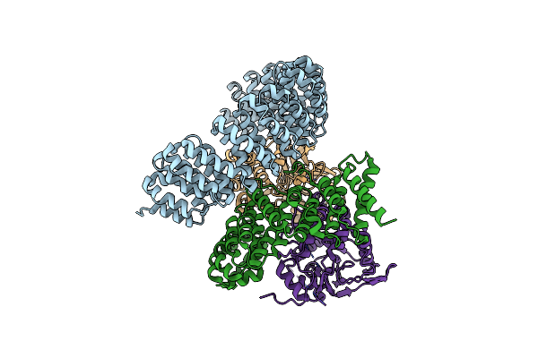
Organism: Mycobacterium tuberculosis h37rv
Method: X-RAY DIFFRACTION Resolution:3.15 Å Release Date: 2024-07-03 Classification: DNA BINDING PROTEIN |
 |
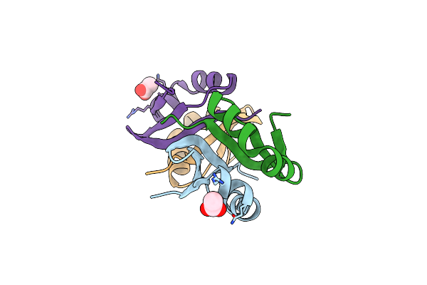
Crystal Structure Of Padr- Family Transcriptional Regulator Rv1176C From Mycobacterium Tuberculosis H37Rv.
Organism: Mycobacterium tuberculosis h37rv
Method: X-RAY DIFFRACTION Resolution:2.94 Å Release Date: 2024-03-13 Classification: DNA BINDING PROTEIN Ligands: GOL, CL, P6G |
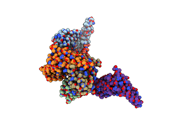
 |
Organism: Homo sapiens
Method: ELECTRON MICROSCOPY Release Date: 2022-08-31 Classification: RECOMBINATION |
 |
Crystal Structure Of N-Terminal Domain Of Vapb46 Antitoxin From Mycobacterium Tuberculosis
Organism: Mycobacterium tuberculosis (strain cdc 1551 / oshkosh)
Method: X-RAY DIFFRACTION Release Date: 2020-04-08 Classification: DNA BINDING PROTEIN Ligands: ACT |
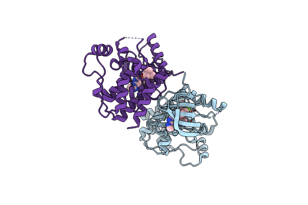 |
Organism: Homo sapiens
Method: X-RAY DIFFRACTION Resolution:2.70 Å Release Date: 2015-07-15 Classification: TRANSFERASE/TRANSFERASE INHIBITOR Ligands: 4Z5, CL |