
Search Count: 16
 |
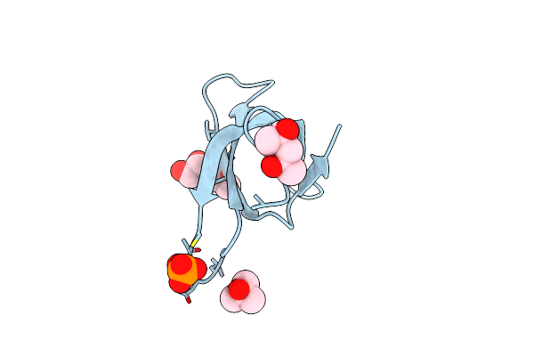
De Novo Designed Bbf-14 Beta Barrel With Computationally Designed Bbf-14_B4 Binder
Organism: Synthetic construct
Method: X-RAY DIFFRACTION Release Date: 2024-12-18 Classification: DE NOVO PROTEIN Ligands: P6G, K, PG4, CL |
 |
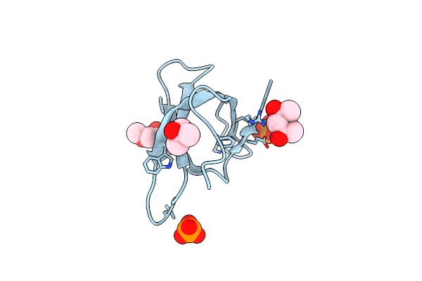
Der F 21 Dust Mite Allergen With Computationally Designed Derf21_B10 Binder
Organism: Dermatophagoides farinae, Synthetic construct
Method: X-RAY DIFFRACTION Release Date: 2024-12-18 Classification: DE NOVO PROTEIN |
 |
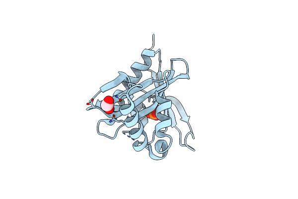
Organism: Dermatophagoides farinae, Synthetic construct
Method: X-RAY DIFFRACTION Release Date: 2024-12-18 Classification: DE NOVO PROTEIN |
 |
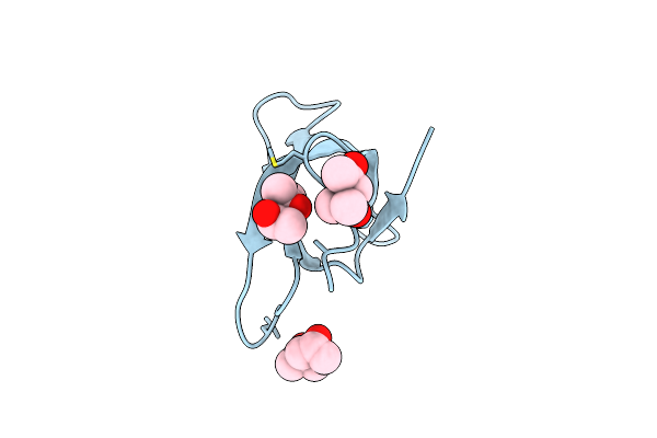
Organism: Dermatophagoides farinae, Synthetic construct
Method: X-RAY DIFFRACTION Release Date: 2024-12-18 Classification: DE NOVO PROTEIN |
 |
Carboxypeptidase G2 Modified With A Versatile Bioconjugate For Metalloprotein Design
Organism: Pseudomonas sp. (strain rs-16)
Method: X-RAY DIFFRACTION Resolution:3.11 Å Release Date: 2021-06-30 Classification: HYDROLASE Ligands: ZN, SO4 |
 |
Crystal Structure Of A Circular Permutation And Computationally Designed Pro-Enzyme Of Carboxypeptidase G2
Organism: Pseudomonas sp. (strain rs-16), Synthetic construct
Method: X-RAY DIFFRACTION Resolution:2.59 Å Release Date: 2021-04-14 Classification: HYDROLASE Ligands: ZN, SO4 |
 |
Crystal Structure Of Trimethoprim-Resistant Type Ii Dihydrofolate Reductase In Complex With A Bisbenzimidazole Inhibitor
Organism: Escherichia coli
Method: X-RAY DIFFRACTION Resolution:1.75 Å Release Date: 2019-05-29 Classification: OXIDOREDUCTASE Ligands: D49, MRD, PO4 |
 |
Crystal Structure Of Trimethoprim-Resistant Type Ii Dihydrofolate Reductase In Complex With A Bisbenzimidazole Inhibitor
Organism: Escherichia coli
Method: X-RAY DIFFRACTION Resolution:1.40 Å Release Date: 2019-05-29 Classification: OXIDOREDUCTASE Ligands: LBA, MRD, PO4 |
 |
Organism: Enterococcus faecium
Method: X-RAY DIFFRACTION Resolution:2.10 Å Release Date: 2016-07-06 Classification: TRANSFERASE Ligands: PO4, EDO |
 |
Epsilon-Caprolactone-Bound Crystal Structure Of Cyclohexanone Monooxygenase In The Tight Conformation
Organism: Rhodococcus sp. hi-31
Method: X-RAY DIFFRACTION Resolution:1.94 Å Release Date: 2014-10-15 Classification: OXIDOREDUCTASE Ligands: FAD, NAP, ECE, BCN |
 |
Epsilon-Caprolactone-Bound Crystal Structure Of Cyclohexanone Monooxygenase In The Loose Conformation
Organism: Rhodococcus sp. hi-31
Method: X-RAY DIFFRACTION Resolution:2.51 Å Release Date: 2014-10-15 Classification: OXIDOREDUCTASE Ligands: FAD, NAP, ECE, PTD |
 |
Cyclohexanone-Bound Crystal Structure Of Cyclohexanone Monooxygenase In The Rotated Conformation
Organism: Rhodococcus sp. hi-31
Method: X-RAY DIFFRACTION Resolution:2.36 Å Release Date: 2012-04-25 Classification: OXIDOREDUCTASE Ligands: FAD, NAP, CYH |
 |
Novel Crystallization Conditions For Tandem Variant R67 Dhfr Yields Wild-Type Crystal Structure
Organism: Escherichia coli
Method: X-RAY DIFFRACTION Resolution:1.40 Å Release Date: 2011-11-02 Classification: OXIDOREDUCTASE Ligands: MRD |
 |
Crystal Structure Of A Methotrexate-Resistant Mutant Of Human Dihydrofolate Reductase
Organism: Homo sapiens
Method: X-RAY DIFFRACTION Resolution:1.70 Å Release Date: 2009-05-26 Classification: OXIDOREDUCTASE Ligands: MTX, SO4, CD |
 |
Organism: Rhodococcus sp.
Method: X-RAY DIFFRACTION Resolution:2.30 Å Release Date: 2009-05-05 Classification: OXIDOREDUCTASE Ligands: FAD, NAP |
 |
Organism: Rhodococcus sp.
Method: X-RAY DIFFRACTION Resolution:2.20 Å Release Date: 2009-05-05 Classification: OXIDOREDUCTASE Ligands: FAD, NAP |