
Search Count: 240
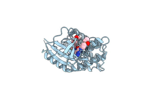 |
Organism: Homo sapiens
Method: X-RAY DIFFRACTION Release Date: 2025-09-24 Classification: CELL CYCLE Ligands: A1D6Z |
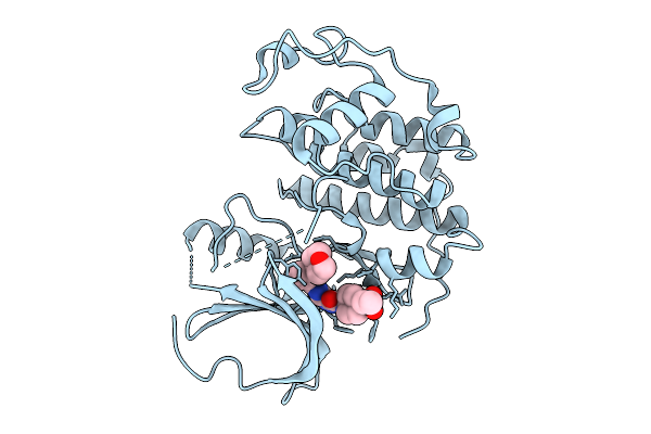 |
Organism: Homo sapiens
Method: X-RAY DIFFRACTION Release Date: 2025-09-24 Classification: CELL CYCLE Ligands: A1D60 |
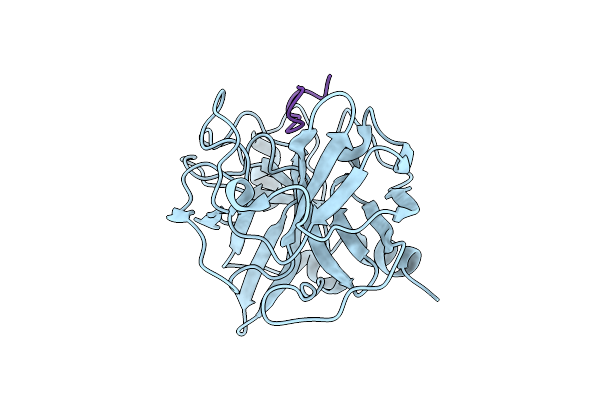 |
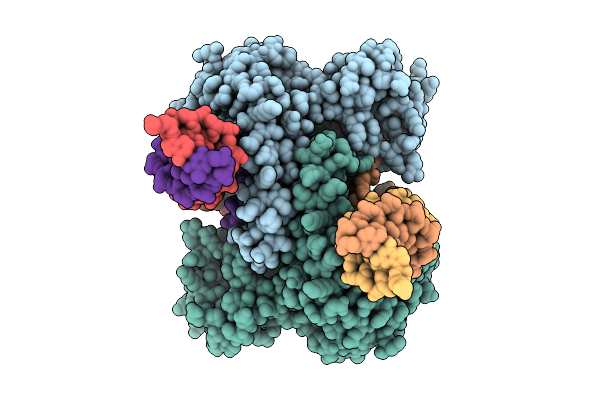
Organism: Homo sapiens, Synthetic construct
Method: X-RAY DIFFRACTION Release Date: 2025-08-06 Classification: PEPTIDE BINDING PROTEIN |
 |
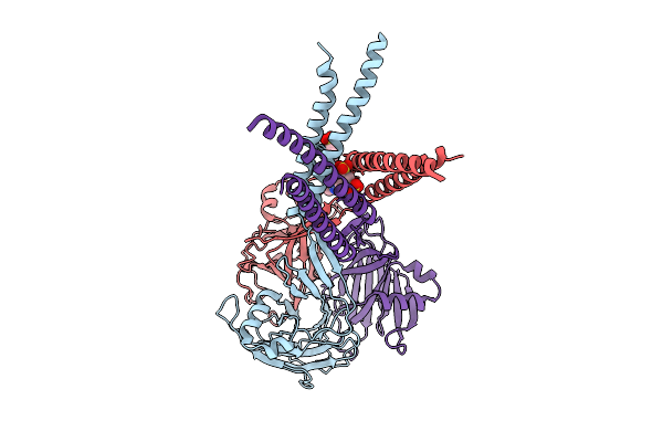
Organism: Homo sapiens
Method: ELECTRON MICROSCOPY Release Date: 2025-07-30 Classification: MEMBRANE PROTEIN |
 |
Organism: Homo sapiens
Method: ELECTRON MICROSCOPY Release Date: 2025-07-30 Classification: MEMBRANE PROTEIN Ligands: DXC |
 |
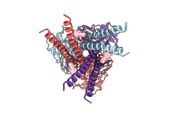
Organism: Homo sapiens
Method: ELECTRON MICROSCOPY Release Date: 2025-07-23 Classification: MEMBRANE PROTEIN Ligands: IZ8 |
 |
Organism: Homo sapiens
Method: ELECTRON MICROSCOPY Release Date: 2025-07-23 Classification: MEMBRANE PROTEIN Ligands: CLR |
 |
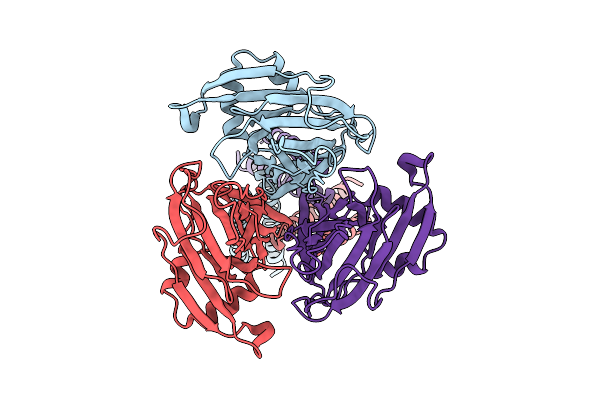
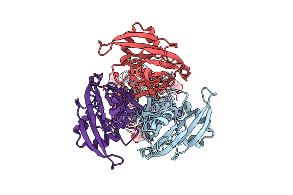
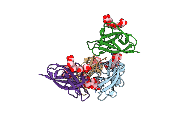
Organism: Homo sapiens
Method: X-RAY DIFFRACTION Release Date: 2025-07-02 Classification: CELL ADHESION |
 |
Organism: Homo sapiens
Method: X-RAY DIFFRACTION Release Date: 2025-07-02 Classification: CELL ADHESION |
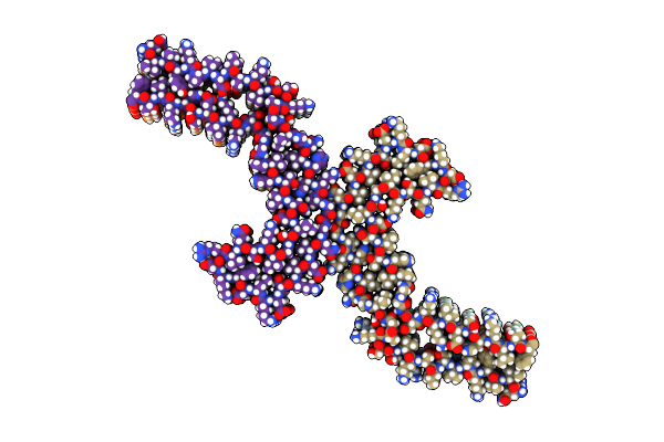 |
Organism: Homo sapiens
Method: ELECTRON MICROSCOPY Release Date: 2025-06-11 Classification: PROTEIN FIBRIL |
 |
Organism: Homo sapiens
Method: ELECTRON MICROSCOPY Release Date: 2025-06-11 Classification: PROTEIN FIBRIL |
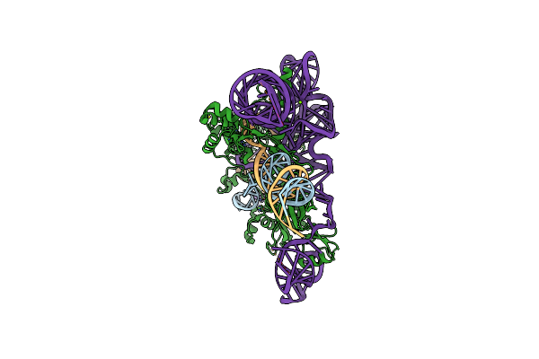 |
Organism: Synthetic construct, Metagenome
Method: ELECTRON MICROSCOPY Release Date: 2025-05-21 Classification: RNA BINDING PROTEIN/RNA/DNA Ligands: MG, ZN |
 |
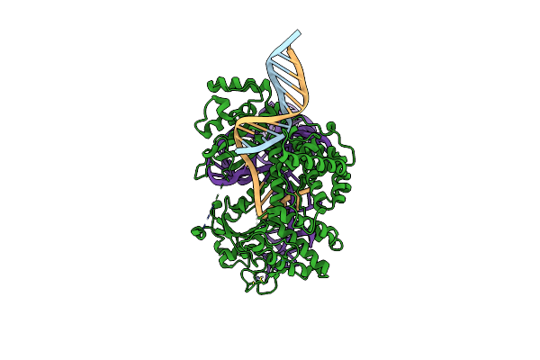
Structure Of Tigr-Tasr In Complex With Tigrna And Target Dna After Dna Cleavage
Organism: Thermoproteota archaeon, Escherichia coli
Method: ELECTRON MICROSCOPY Resolution:3.05 Å Release Date: 2025-03-05 Classification: DNA BINDING PROTEIN/RNA/DNA Ligands: MG |
 |
Organism: Homo sapiens
Method: ELECTRON MICROSCOPY Release Date: 2024-10-09 Classification: HYDROLASE Ligands: GDP, ANP, MG, GNP |
 |
Organism: Homo sapiens
Method: ELECTRON MICROSCOPY Release Date: 2024-10-09 Classification: HYDROLASE Ligands: GDP, ANP, MG, GNP |
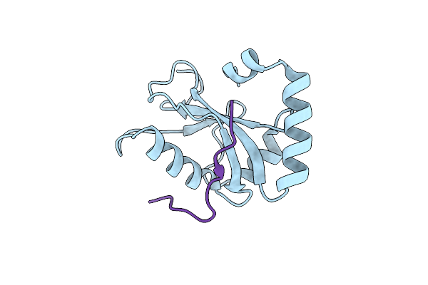 |
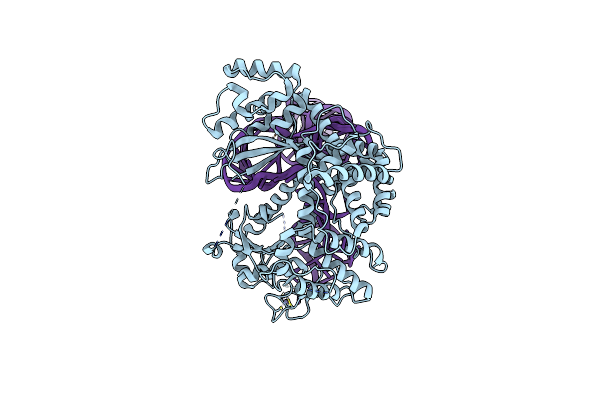
Organism: Homo sapiens
Method: X-RAY DIFFRACTION Resolution:1.53 Å Release Date: 2024-09-18 Classification: PROTEIN BINDING |
 |
Organism: Homo sapiens
Method: X-RAY DIFFRACTION Resolution:2.10 Å Release Date: 2024-09-18 Classification: PROTEIN BINDING Ligands: GOL |
 |
Organism: Escherichia coli k-12, Guillardia theta
Method: ELECTRON MICROSCOPY Release Date: 2024-09-11 Classification: RNA BINDING PROTEIN/RNA/DNA Ligands: ZN |
 |
Organism: Escherichia coli k-12, Guillardia theta, Synthetic construct
Method: ELECTRON MICROSCOPY Release Date: 2024-09-11 Classification: RNA BINDING PROTEIN/RNA/DNA Ligands: ZN |
 |
Organism: Escherichia coli k-12, Guillardia theta, Synthetic construct
Method: ELECTRON MICROSCOPY Release Date: 2024-09-11 Classification: RNA BINDING PROTEIN/RNA/DNA Ligands: ZN |