
Search Count: 263
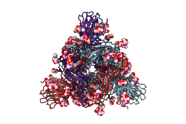 |
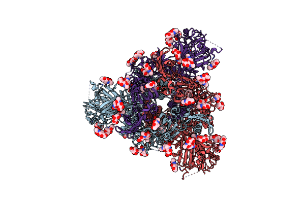
Organism: Sarbecovirus
Method: ELECTRON MICROSCOPY Release Date: 2025-10-29 Classification: VIRAL PROTEIN Ligands: BLA, NAG, EIC |
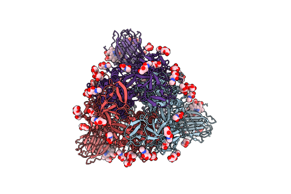 |
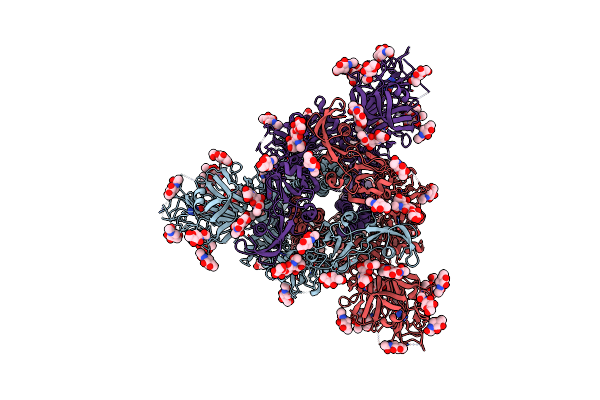
Organism: Sarbecovirus
Method: ELECTRON MICROSCOPY Release Date: 2025-10-29 Classification: VIRAL PROTEIN Ligands: NAG, BLA |
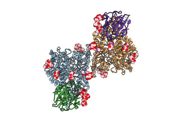 |
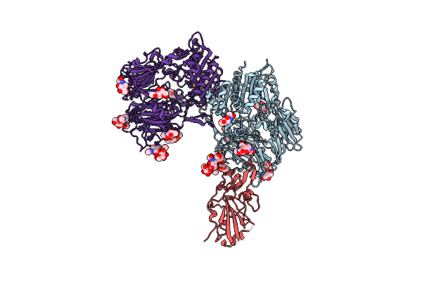
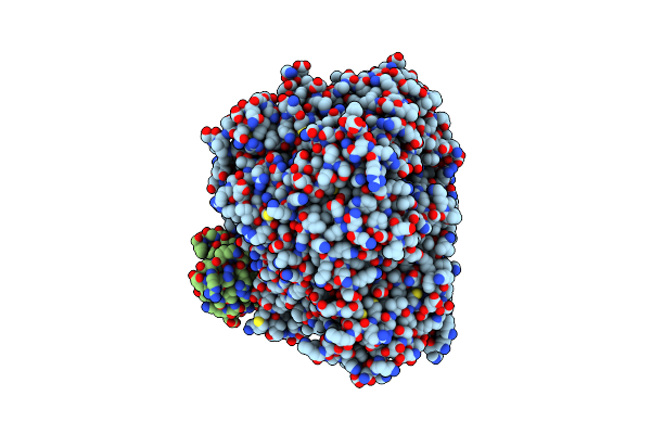
Rhinolophus Cornutus Bat Ace2 Dimer In Complex With Two Rc-O319 Sarbecovirus Spike Rbds.
Organism: Sarbecovirus, Rhinolophus cornutus
Method: ELECTRON MICROSCOPY Release Date: 2025-10-29 Classification: VIRAL PROTEIN/HYDROLASE Ligands: NAG |
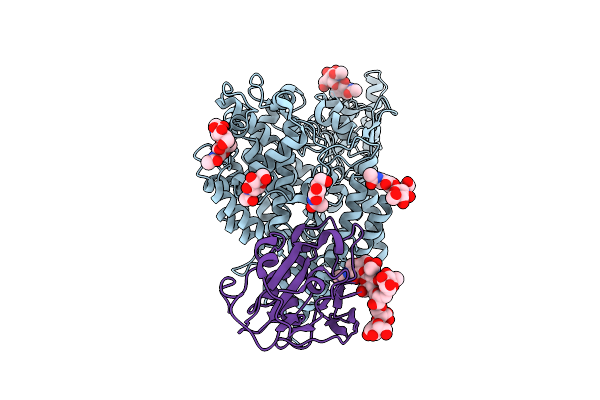 |
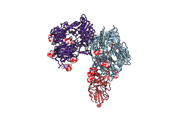
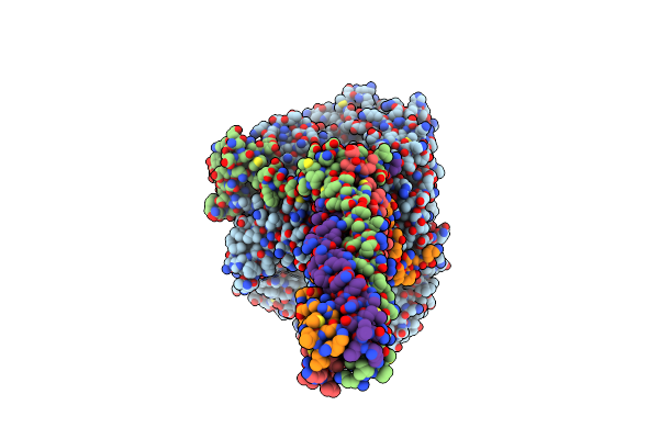
Rhinolophus Cornutus Bat Ace2 Dimer In Complex With Two Rc-O319 Sarbecovirus Spike Rbds.
Organism: Sarbecovirus, Rhinolophus cornutus
Method: ELECTRON MICROSCOPY Release Date: 2025-10-29 Classification: VIRAL PROTEIN/HYDROLASE Ligands: NAG |
 |
Organism: Coronavirus neoromicia/pml-phe1/rsa/2011, Enterobacteria phage t4
Method: ELECTRON MICROSCOPY Release Date: 2025-06-18 Classification: VIRAL PROTEIN Ligands: NAG, EIC |
 |
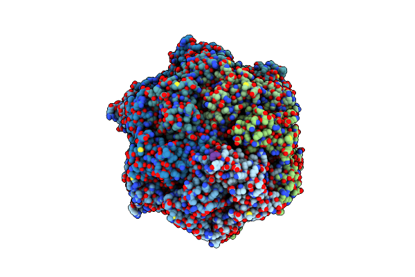
Cryo-Em Structure Of Eu-Hedgehogcov (Erinaceus/Vmc/Deu/2012) S-Trimer In A Locked-2 Conformation
Organism: Betacoronavirus erinaceus/vmc/deu/2012, Enterobacteria phage t4 (bacteriophage t4)
Method: ELECTRON MICROSCOPY Release Date: 2025-06-18 Classification: VIRAL PROTEIN Ligands: NAG, FOL, EIC |
 |
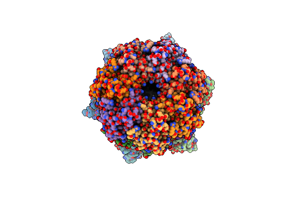
Cryo-Em Structure Of Hku25-Batcov S-Trimer Stabilized With 2P And X1 Disulfide Bond
Organism: Hypsugo bat coronavirus hku25, Tequatrovirus t4
Method: ELECTRON MICROSCOPY Release Date: 2025-06-18 Classification: VIRAL PROTEIN Ligands: EIC, NAG |
 |
Cryo-Em Structure Of Cn-Hedgehogcov (Hku31/Erinaceus Amurensis/China/2014) S-Trimer In A Locked-2 Conformation
Organism: Erinaceus hedgehog coronavirus hku31, Enterobacteria phage t4
Method: ELECTRON MICROSCOPY Release Date: 2025-06-18 Classification: VIRAL PROTEIN Ligands: FOL, NAG |
 |
Cryo-Em Structure Of The Gd-Batcov (Btcov/Ii/Gd/2014-422) Rbd In Complex With Human Dpp4
Organism: Homo sapiens, Middle east respiratory syndrome-related coronavirus
Method: ELECTRON MICROSCOPY Release Date: 2025-06-18 Classification: VIRAL PROTEIN Ligands: NAG |
 |
Cryo-Em Structure Of The Se-Pangolincov (Mjhku4R-Cov-1) Rbd In Complex With Human Dpp4
Organism: Homo sapiens, Pangolin coronavirus hku4/p251t/pangolin/2018
Method: ELECTRON MICROSCOPY Release Date: 2025-06-18 Classification: VIRAL PROTEIN Ligands: NAG |
 |
Cryo-Em Structure Of Cn-Hedgehogcov (Hku31/Erinaceus Amurensis/China/2014) S-Trimer In A Locked-1 Conformation
Organism: Erinaceus hedgehog coronavirus hku31, Tequatrovirus t4
Method: ELECTRON MICROSCOPY Release Date: 2025-06-18 Classification: VIRAL PROTEIN Ligands: NAG, FOL, EIC |
 |
Organism: Bat coronavirus, Tequatrovirus t4
Method: ELECTRON MICROSCOPY Release Date: 2025-06-18 Classification: VIRAL PROTEIN Ligands: NAG, EIC |
 |
Organism: Middle east respiratory syndrome-related coronavirus, Tequatrovirus t4
Method: ELECTRON MICROSCOPY Release Date: 2025-06-18 Classification: VIRAL PROTEIN Ligands: NAG, EIC |
 |
Cryo-Em Structure Of A Truncated Nipah Virus L Protein Bound By Phosphoprotein Tetramer
Organism: Henipavirus nipahense
Method: ELECTRON MICROSCOPY Release Date: 2025-05-21 Classification: VIRAL PROTEIN Ligands: ZN |
 |
Cryo-Em Structure Of The Full-Length Nipah Virus L Protein Bound By Phosphoprotein Tetramer
Organism: Henipavirus nipahense
Method: ELECTRON MICROSCOPY Release Date: 2025-05-21 Classification: VIRAL PROTEIN Ligands: ZN |
 |
Cryoem Structure Of M. Tuberculosis Clpc1P1P2 Complex Bound To Bortezomib, Conformation 2
Organism: Mycobacterium tuberculosis h37rv, Bos grunniens
Method: ELECTRON MICROSCOPY Release Date: 2025-03-19 Classification: HYDROLASE Ligands: ADP, ATP, MG, BO2 |
 |
Organism: Mycobacterium tuberculosis h37rv
Method: ELECTRON MICROSCOPY Release Date: 2025-03-19 Classification: HYDROLASE Ligands: MG, ATP, ADP, BO2 |
 |
Cryoem Structure Of M. Tuberculosis Clpc1P1P2 Complex Bound To Bortezomib, Conformation 1
Organism: Mycobacterium tuberculosis h37rv, Bos grunniens
Method: ELECTRON MICROSCOPY Release Date: 2025-03-19 Classification: HYDROLASE Ligands: MG, ATP, ADP, BO2 |
 |
Organism: Mycobacterium tuberculosis h37rv
Method: ELECTRON MICROSCOPY Release Date: 2025-03-19 Classification: HYDROLASE Ligands: BO2 |
 |
Cryoem Structure Of M. Tuberculosis Clpc1P1P2 Complex Bound To Bortezomib, Conformation 3
Organism: Mycobacterium tuberculosis h37rv, Bos grunniens
Method: ELECTRON MICROSCOPY Release Date: 2025-03-19 Classification: HYDROLASE Ligands: ATP, MG, ADP, BO2 |