
Search Count: 62
 |
Organism: Escherichia phage t4, Homo sapiens
Method: ELECTRON MICROSCOPY Release Date: 2025-10-22 Classification: ISOMERASE Ligands: MG |
 |
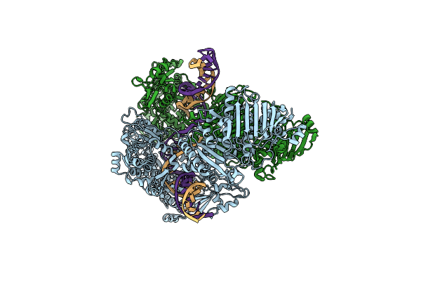
Organism: Escherichia phage t4, Dna molecule
Method: ELECTRON MICROSCOPY Release Date: 2025-10-01 Classification: ISOMERASE/DNA |
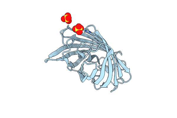 |
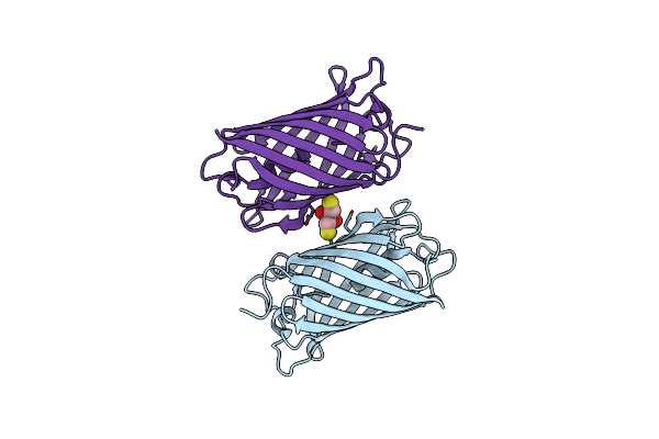
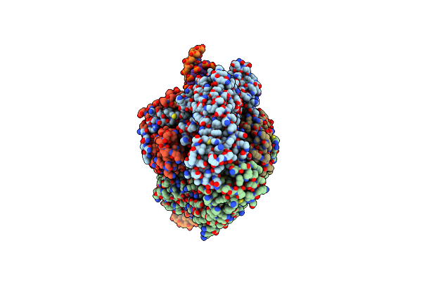
Organism: Lobophyllia hemprichii
Method: X-RAY DIFFRACTION Resolution:1.34 Å Release Date: 2025-04-16 Classification: FLUORESCENT PROTEIN Ligands: SO4 |
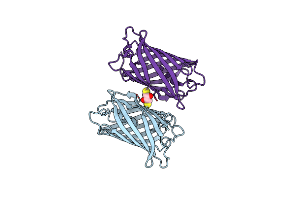 |
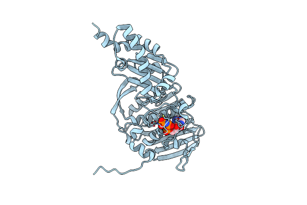
Organism: Lobophyllia hemprichii
Method: X-RAY DIFFRACTION Resolution:1.66 Å Release Date: 2025-04-16 Classification: FLUORESCENT PROTEIN Ligands: DTT |
 |
Organism: Lobophyllia hemprichii
Method: X-RAY DIFFRACTION Resolution:1.85 Å Release Date: 2025-04-16 Classification: FLUORESCENT PROTEIN Ligands: DTT |
 |
Organism: Unidentified, Unclassified sequences
Method: ELECTRON MICROSCOPY Release Date: 2024-12-11 Classification: RNA BINDING PROTEIN/RNA/DNA |
 |
Organism: Unclassified sequences
Method: ELECTRON MICROSCOPY Release Date: 2024-12-11 Classification: RNA BINDING PROTEIN/RNA/DNA |
 |
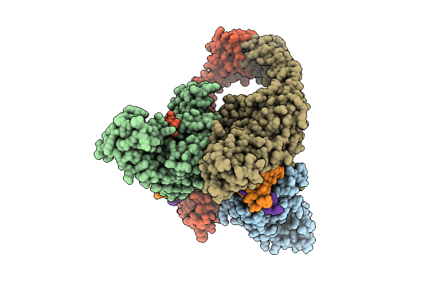
Organism: Escherichia phage t4, Salmonella phage chi, Dna molecule
Method: ELECTRON MICROSCOPY Release Date: 2024-09-25 Classification: ISOMERASE Ligands: MG |
 |
Organism: Enterobacteria phage t6
Method: ELECTRON MICROSCOPY Release Date: 2024-09-25 Classification: ISOMERASE Ligands: ANP, MG |
 |
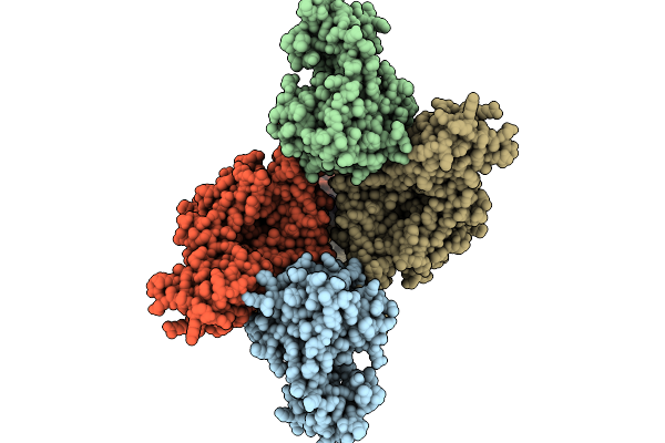
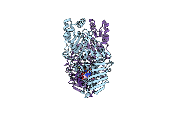
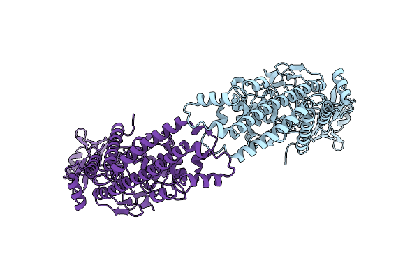
Organism: Escherichia phage t4
Method: ELECTRON MICROSCOPY Release Date: 2024-09-25 Classification: ISOMERASE |
 |
Organism: Escherichia phage t4, Dna molecule
Method: ELECTRON MICROSCOPY Release Date: 2024-09-25 Classification: ISOMERASE Ligands: MG |
 |
Organism: Escherichia phage t4, Enterobacteria phage t6
Method: ELECTRON MICROSCOPY Release Date: 2024-09-25 Classification: ISOMERASE |
 |
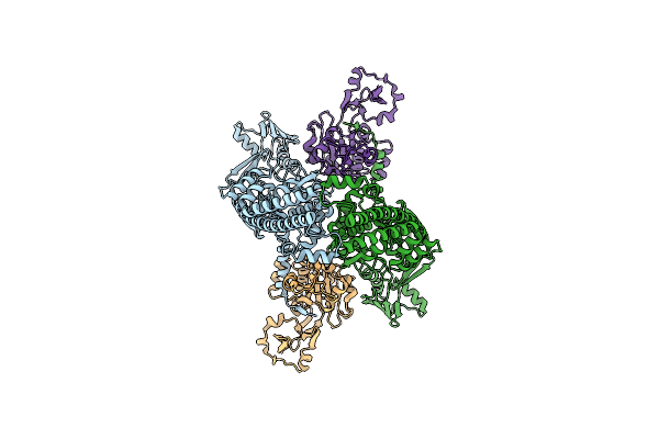
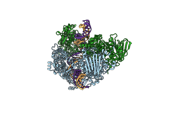
Structure Of Phage T6 Topoisomerase Ii Central Domain Bound With Dna And M-Amsa
Organism: Escherichia phage t4, Enterobacteria phage t6, Dna molecule
Method: ELECTRON MICROSCOPY Release Date: 2024-09-25 Classification: ISOMERASE Ligands: MG, ASW |
 |
Organism: Escherichia phage t4
Method: ELECTRON MICROSCOPY Release Date: 2024-09-25 Classification: ISOMERASE |
 |
Organism: Escherichia phage t4, Enterobacteria phage t6
Method: ELECTRON MICROSCOPY Release Date: 2024-09-25 Classification: ISOMERASE Ligands: ANP |
 |
Organism: Escherichia phage t4, Enterobacteria phage t6, Dna molecule
Method: ELECTRON MICROSCOPY Release Date: 2024-09-25 Classification: ISOMERASE Ligands: ANP |
 |
Organism: Enterobacteria phage t6
Method: X-RAY DIFFRACTION Resolution:2.80 Å Release Date: 2024-09-25 Classification: ISOMERASE Ligands: MG, ANP |
 |
Organism: African swine fever virus lis57
Method: ELECTRON MICROSCOPY Release Date: 2024-04-03 Classification: VIRAL PROTEIN |
 |
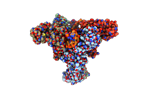
Structure Of African Swine Fever Virus Topoisomerase Ii In Complex With Dsdna
Organism: African swine fever virus, Dna molecule
Method: ELECTRON MICROSCOPY Release Date: 2024-04-03 Classification: VIRAL PROTEIN/DNA |
 |
Structure Of African Swine Fever Virus Topoisomerase Ii In Complex With Dsdna
Organism: African swine fever virus, Dna molecule
Method: ELECTRON MICROSCOPY Release Date: 2024-04-03 Classification: VIRAL PROTEIN/DNA |