
Search Count: 278
 |
Organism: Canis lupus familiaris
Method: X-RAY DIFFRACTION Release Date: 2025-10-29 Classification: CYTOKINE/IMMUNE SYSTEM |
 |
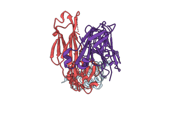
Structural Insights Into Il-31 Signaling Inhibition By A Neutralization Antibody
Organism: Canis lupus familiaris
Method: X-RAY DIFFRACTION Release Date: 2025-10-29 Classification: CYTOKINE/IMMUNE SYSTEM |
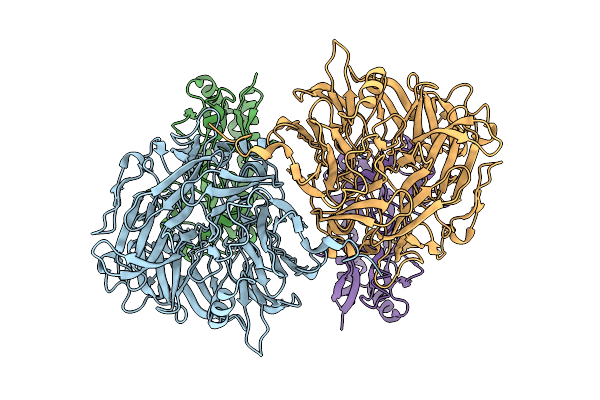 |
Organism: Methylorubrum extorquens
Method: ELECTRON MICROSCOPY Release Date: 2025-07-23 Classification: PROTEIN BINDING |
 |
Organism: Methylorubrum extorquens
Method: ELECTRON MICROSCOPY Release Date: 2025-07-23 Classification: PROTEIN BINDING Ligands: PQQ |
 |
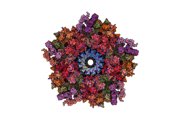
Organism: Bovine adenovirus 3
Method: ELECTRON MICROSCOPY Release Date: 2025-06-04 Classification: VIRAL PROTEIN |
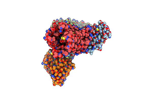 |
Organism: Homo sapiens, Mus musculus
Method: ELECTRON MICROSCOPY Release Date: 2025-04-23 Classification: MEMBRANE PROTEIN/IMMUNE SYSTEM Ligands: A1D5N |
 |
Organism: Streptomyces lasalocidi
Method: X-RAY DIFFRACTION Resolution:1.85 Å Release Date: 2025-04-16 Classification: FLAVOPROTEIN Ligands: FAD, GOL, CL |
 |
Organism: Escherichia phage n4
Method: ELECTRON MICROSCOPY Release Date: 2025-04-16 Classification: VIRAL PROTEIN |
 |
Organism: Enterobacteria phage n4
Method: ELECTRON MICROSCOPY Release Date: 2025-04-16 Classification: VIRAL PROTEIN |
 |
Organism: Enterobacteria phage n4
Method: ELECTRON MICROSCOPY Release Date: 2025-04-16 Classification: VIRAL PROTEIN |
 |
Organism: Enterobacteria phage n4
Method: ELECTRON MICROSCOPY Release Date: 2025-04-16 Classification: VIRUS |
 |
Organism: Streptomyces virginiae
Method: X-RAY DIFFRACTION Resolution:2.00 Å Release Date: 2025-04-16 Classification: FLAVOPROTEIN Ligands: FAD, Y7R, CL |
 |
Organism: Streptomyces virginiae
Method: X-RAY DIFFRACTION Resolution:2.10 Å Release Date: 2025-04-16 Classification: FLAVOPROTEIN Ligands: FAD, GOL, YGK, CL |
 |
Organism: Streptomyces virginiae
Method: X-RAY DIFFRACTION Resolution:2.65 Å Release Date: 2025-04-16 Classification: FLAVOPROTEIN Ligands: FAD, YK6, GOL, CL |
 |
Organism: Homo sapiens
Method: X-RAY DIFFRACTION Resolution:2.10 Å Release Date: 2025-04-02 Classification: STRUCTURAL PROTEIN Ligands: A1EKR, GOL |
 |
Organism: Homo sapiens
Method: X-RAY DIFFRACTION Resolution:2.33 Å Release Date: 2025-04-02 Classification: SIGNALING PROTEIN Ligands: A1EKR |
 |
Organism: Escherichia phage t1
Method: ELECTRON MICROSCOPY Release Date: 2025-03-12 Classification: VIRAL PROTEIN |
 |
Organism: Escherichia phage t1
Method: ELECTRON MICROSCOPY Release Date: 2025-03-12 Classification: VIRAL PROTEIN |
 |
Organism: Escherichia phage t1
Method: ELECTRON MICROSCOPY Release Date: 2025-03-12 Classification: VIRAL PROTEIN |
 |
Organism: Escherichia phage t1
Method: ELECTRON MICROSCOPY Release Date: 2025-03-12 Classification: VIRAL PROTEIN |