
Search Count: 35
 |
Organism: Tetrahymena thermophila
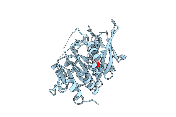
Method: X-RAY DIFFRACTION Release Date: 2025-11-12 Classification: HYDROLASE Ligands: GOL |
 |
Organism: Tetrahymena thermophila
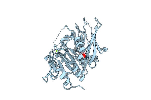
Method: X-RAY DIFFRACTION Release Date: 2025-11-12 Classification: HYDROLASE Ligands: MG, GOL |
 |
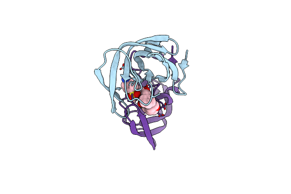
Organism: Tetrahymena thermophila
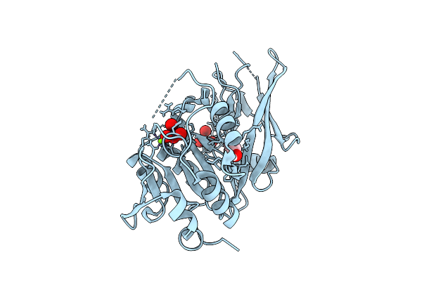
Method: X-RAY DIFFRACTION Release Date: 2025-11-12 Classification: HYDROLASE Ligands: WO4, MG, GOL |
 |
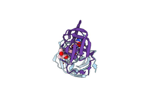
Organism: Tetrahymena thermophila
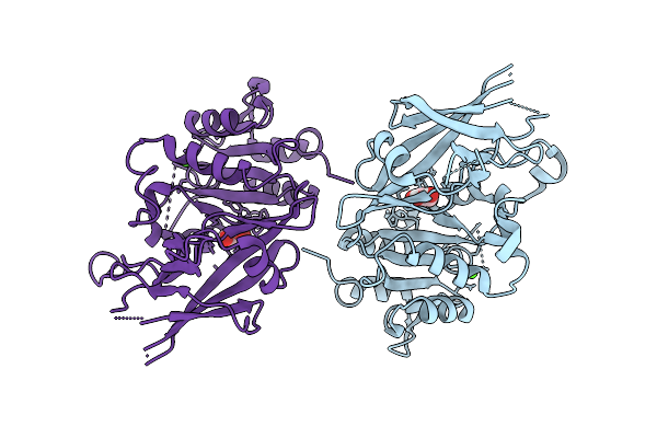
Method: X-RAY DIFFRACTION Release Date: 2025-11-12 Classification: HYDROLASE Ligands: CA, GOL |
 |
Organism: Emericella nidulans (strain fgsc a4 / atcc 38163 / cbs 112.46 / nrrl 194 / m139)
Method: X-RAY DIFFRACTION Resolution:2.09 Å Release Date: 2022-04-27 Classification: FLAVOPROTEIN Ligands: FAD, SO4 |
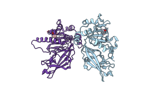 |
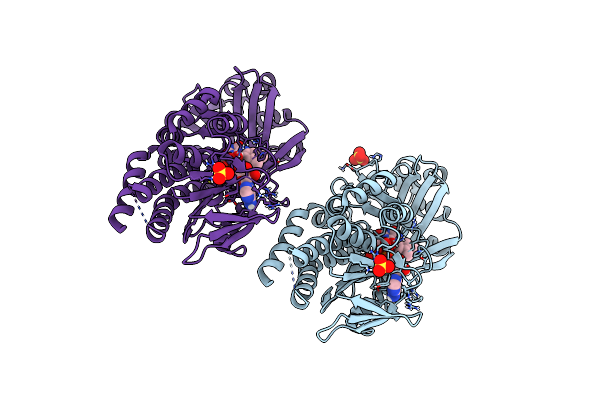
High-Resolution Crystal Structure Of A Lipin/Pah Phosphatidic Acid Phosphatase
Organism: Tetrahymena thermophila (strain sb210)
Method: X-RAY DIFFRACTION Release Date: 2022-02-23 Classification: HYDROLASE Ligands: CAC |
 |
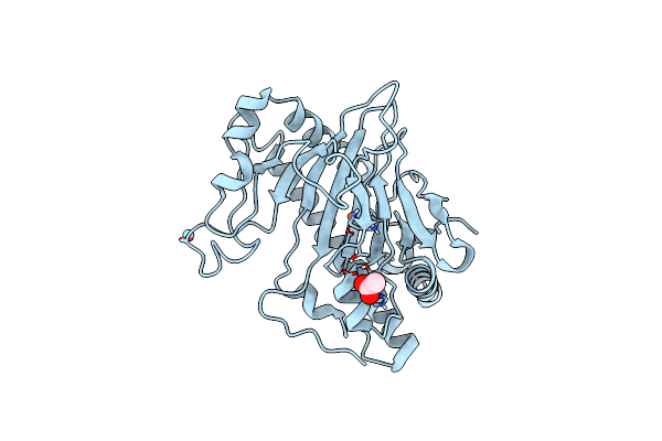
X-Ray Crystal Structure Of E.Coli Dihydrofolate Reductase Complexed With Folate And Nadp+ At Ph4.5
Organism: Escherichia coli (strain k12)
Method: X-RAY DIFFRACTION Resolution:1.65 Å Release Date: 2021-06-09 Classification: OXIDOREDUCTASE Ligands: FOL, NAP |
 |
X-Ray Crystal Structure Of E.Coli Dihydrofolate Reductase Complexed With Folate And Nadp+ At Ph4.5
Organism: Escherichia coli (strain k12)
Method: X-RAY DIFFRACTION Resolution:1.65 Å Release Date: 2021-06-09 Classification: OXIDOREDUCTASE Ligands: FOL, NAP |
 |
X-Ray Crystal Structure Of E.Coli Dihydrofolate Reductase Complexed With Folate And Nadp+ At Ph7.0
Organism: Escherichia coli (strain k12)
Method: X-RAY DIFFRACTION Resolution:1.60 Å Release Date: 2021-06-09 Classification: OXIDOREDUCTASE Ligands: FOL, NAP, MN |
 |
X-Ray Crystal Structure Of E.Coli Dihydrofolate Reductase Complexed With Folate And Nadp+ At Ph7.0
Organism: Escherichia coli (strain k12)
Method: X-RAY DIFFRACTION Resolution:1.60 Å Release Date: 2021-06-09 Classification: OXIDOREDUCTASE Ligands: FOL, NAP |
 |
Neutron Crystal Structure Of E.Coli Dihydrofolate Reductase Complexed With Folate And Nadp+ At Ph4.5
Organism: Escherichia coli (strain k12)
Method: X-RAY DIFFRACTION, NEUTRON DIFFRACTION Resolution:1.6500 Å, 2.1 Å Release Date: 2021-06-09 Classification: OXIDOREDUCTASE Ligands: MN, FOL, NAP, DOD |
 |
Organism: Talaromyces stipitatus (strain atcc 10500 / cbs 375.48 / qm 6759 / nrrl 1006)
Method: X-RAY DIFFRACTION Resolution:2.70 Å Release Date: 2021-06-02 Classification: OXIDOREDUCTASE Ligands: FE, YT3, ACT |
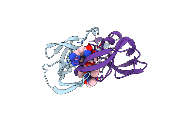 |
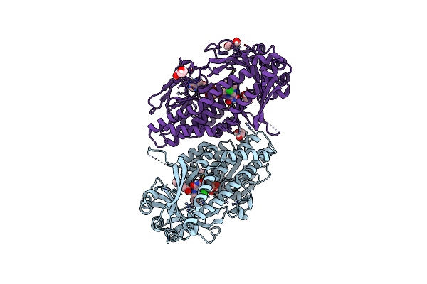
100K X-Ray Structure Of Hiv-1 Protease Triple Mutant (V32I,I47V,V82I) With Tetrahedral Intermediate Mimic Kvs-1
Organism: Human immunodeficiency virus 1
Method: X-RAY DIFFRACTION Resolution:1.31 Å Release Date: 2020-07-29 Classification: VIRAL PROTEIN Ligands: KVS |
 |
Room Temperature X-Ray Structure Of Hiv-1 Protease Triple Mutant (V32I,I47V,V82I) With Tetrahedral Intermediate Of Keto-Darunavir
Organism: Human immunodeficiency virus 1
Method: X-RAY DIFFRACTION Resolution:1.80 Å Release Date: 2020-06-24 Classification: HYDROLASE Ligands: P3V |
 |
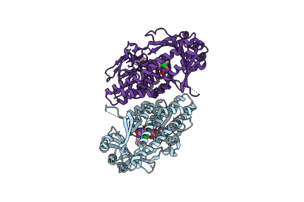
Joint X-Ray/Neutron Structure Of Hiv-1 Protease Triple Mutant (V32I,I47V,V82I) With Tetrahedral Intermediate Mimic Kvs-1
Organism: Human immunodeficiency virus 1
Method: X-RAY DIFFRACTION, NEUTRON DIFFRACTION Resolution:1.8500 Å, 2.2000 Å Release Date: 2020-06-10 Classification: HYDROLASE Ligands: KVS, DOD |
 |
Organism: Talaromyces stipitatus (strain atcc 10500 / cbs 375.48 / qm 6759 / nrrl 1006)
Method: X-RAY DIFFRACTION Resolution:1.75 Å Release Date: 2019-08-14 Classification: FLAVOPROTEIN Ligands: HEZ, GOL, FAD, CL |
 |
Organism: Talaromyces stipitatus (strain atcc 10500 / cbs 375.48 / qm 6759 / nrrl 1006)
Method: X-RAY DIFFRACTION Resolution:2.25 Å Release Date: 2019-08-14 Classification: FLAVOPROTEIN Ligands: KJY, FAD, CL, GOL |
 |
Organism: Talaromyces stipitatus (strain atcc 10500 / cbs 375.48 / qm 6759 / nrrl 1006)
Method: X-RAY DIFFRACTION Resolution:2.30 Å Release Date: 2019-08-14 Classification: FLAVOPROTEIN Ligands: FAD, CL, GOL |
 |
Organism: Talaromyces stipitatus (strain atcc 10500 / cbs 375.48 / qm 6759 / nrrl 1006)
Method: X-RAY DIFFRACTION Resolution:2.30 Å Release Date: 2019-08-14 Classification: FLAVOPROTEIN Ligands: FAD, CL |
 |
Room Temperature Structure Of Binary Complex Of Native Hache With Oxime Reactivator Rs-170B
Organism: Homo sapiens
Method: X-RAY DIFFRACTION Resolution:2.80 Å Release Date: 2019-05-29 Classification: HYDROLASE Ligands: LND, CL |