
Search Count: 17
 |
Organism: Homo sapiens, Clostridium pasteurianum
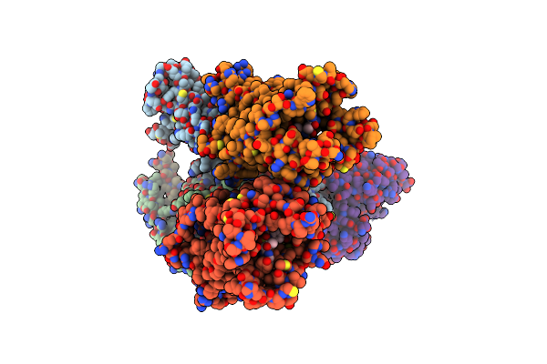
Method: X-RAY DIFFRACTION Resolution:2.70 Å Release Date: 2022-07-27 Classification: MEMBRANE PROTEIN Ligands: ZN, 8EH, OLC |
 |
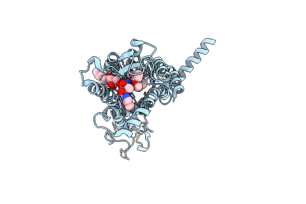
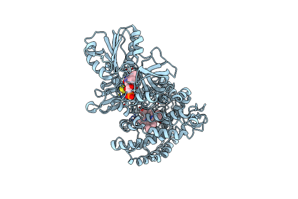
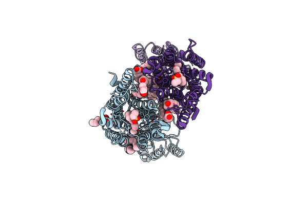
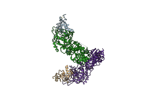
Organism: Homo sapiens, Escherichia coli
Method: ELECTRON MICROSCOPY Release Date: 2022-07-27 Classification: MEMBRANE PROTEIN Ligands: 8EH |
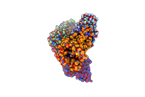 |
Organism: Homo sapiens, Escherichia coli
Method: ELECTRON MICROSCOPY Release Date: 2022-07-27 Classification: MEMBRANE PROTEIN Ligands: 8EH |
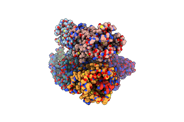 |
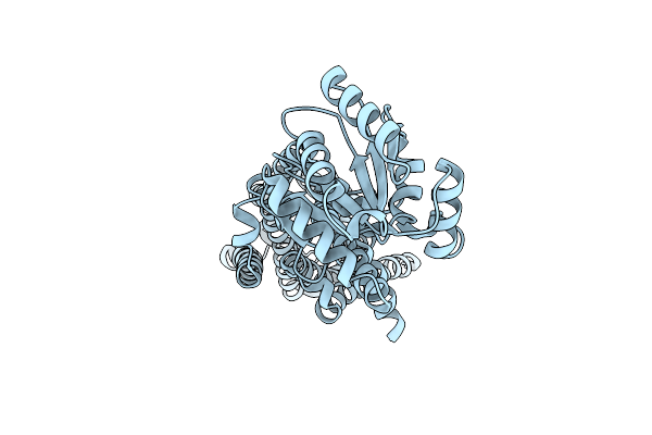
Organism: Homo sapiens, Escherichia coli
Method: ELECTRON MICROSCOPY Release Date: 2022-07-27 Classification: MEMBRANE PROTEIN |
 |
Organism: Homo sapiens, Escherichia coli
Method: ELECTRON MICROSCOPY Release Date: 2022-07-27 Classification: MEMBRANE PROTEIN |
 |
Organism: Homo sapiens, Escherichia coli
Method: ELECTRON MICROSCOPY Release Date: 2022-07-27 Classification: MEMBRANE PROTEIN |
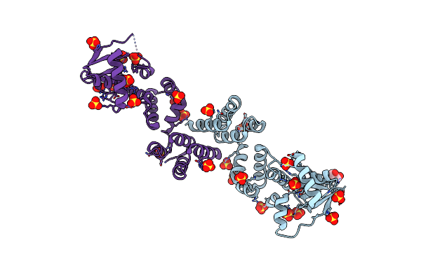 |
Crystal Structure Of C-Terminally Truncated Geobacillus Thermoleovorans Nucleoid Occlusion Protein Noc
Organism: Geobacillus thermoleovorans ccb_us3_uf5
Method: X-RAY DIFFRACTION Resolution:2.50 Å Release Date: 2021-02-17 Classification: DNA BINDING PROTEIN Ligands: SO4, GOL |
 |
Crystal Structure Of N- And C-Terminally Truncated Geobacillus Thermoleovorans Nucleoid Occlusion Protein Noc
Organism: Geobacillus thermoleovorans ccb_us3_uf5
Method: X-RAY DIFFRACTION Resolution:2.95 Å Release Date: 2021-02-17 Classification: DNA BINDING PROTEIN Ligands: SO4 |
 |
Crystal Structure Of An Intact Type Iv Self-Sufficient Cytochrome P450 Monooxygenase
Organism: Rhodococcus sp. ecu0066
Method: X-RAY DIFFRACTION Resolution:2.60 Å Release Date: 2020-07-01 Classification: OXIDOREDUCTASE Ligands: FMN, HEM, FES |
 |
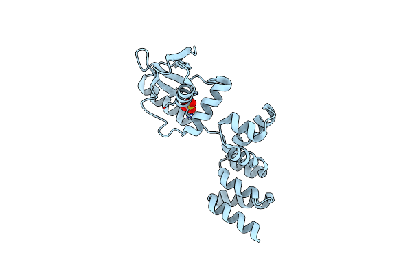
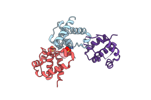
The Structural Study Of Mutation Induced Inactivation Of Human Muscarinic Receptor M4
Organism: Homo sapiens, Pyrococcus abyssi ge5
Method: X-RAY DIFFRACTION Resolution:3.00 Å Release Date: 2020-03-11 Classification: MEMBRANE PROTEIN |
 |
Organism: Homo sapiens
Method: X-RAY DIFFRACTION Resolution:2.80 Å Release Date: 2019-12-04 Classification: SIGNALING PROTEIN Ligands: E33, CLR, OLC, OLA |
 |
X-Ray Structure Of The Apo Form Of The Establishement Gene Regulator Reg576 Of The G+ Plasmid P576
Organism: Bacillus altitudinis
Method: X-RAY DIFFRACTION Resolution:1.98 Å Release Date: 2018-10-17 Classification: TRANSCRIPTION |
 |
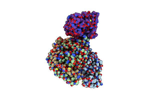
Crystal Structure Of The Sars Coronavirus Nsp14-Nsp10 Complex With Functional Ligands Sah And Gpppa
Organism: Human sars coronavirus
Method: X-RAY DIFFRACTION Resolution:3.33 Å Release Date: 2015-07-15 Classification: TRANSFERASE Ligands: ZN, MG, SAH, G3A |
 |
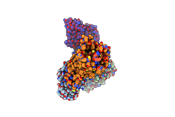
Crystal Structure Of The Sars Coronavirus Nsp14-Nsp10 Complex With Functional Ligand Sam
Organism: Human sars coronavirus
Method: X-RAY DIFFRACTION Resolution:3.20 Å Release Date: 2015-07-15 Classification: TRANSFERASE Ligands: ZN, MG, SAM |
 |
Organism: Human sars coronavirus
Method: X-RAY DIFFRACTION Resolution:3.40 Å Release Date: 2015-07-15 Classification: TRANSFERASE Ligands: ZN, MG |
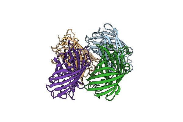 |
Organism: Lobophyllia hemprichii
Method: X-RAY DIFFRACTION Resolution:2.20 Å Release Date: 2011-11-16 Classification: FLUORESCENT PROTEIN |
 |
Organism: Homo sapiens
Method: X-RAY DIFFRACTION Resolution:2.50 Å Release Date: 2011-03-09 Classification: DNA BINDING PROTEIN Ligands: MES, SO4 |