
Search Count: 62
 |
Organism: Axinella sp. 1 tf-2017
Method: SOLUTION NMR Release Date: 2024-09-25 Classification: TOXIN Ligands: PCA |
 |
Structure Of The Laspartomycin C Double Mutant G4D D-Allo-Thr9D-Dap In Complex With Geranyl Phosphate
Organism: Streptomyces viridochromogenes
Method: X-RAY DIFFRACTION Resolution:1.04 Å Release Date: 2022-02-02 Classification: ANTIBIOTIC Ligands: CA, RDZ, 9GE |
 |
Structure Of The Laspartomycin C Friulimicin-Like Mutant In Complex With Geranyl Phosphate
Organism: Streptomyces viridochromogenes
Method: X-RAY DIFFRACTION Resolution:1.14 Å Release Date: 2022-02-02 Classification: ANTIBIOTIC Ligands: CA, RDZ, CL, CD, NA |
 |
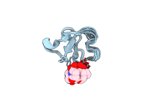
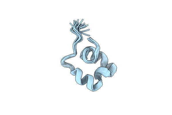
Organism: Necator americanus
Method: SOLUTION NMR Release Date: 2021-10-27 Classification: IMMUNE SYSTEM |
 |
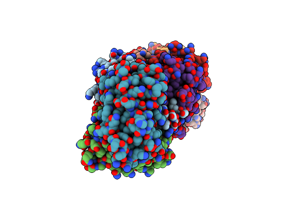
Organism: Shewanella oneidensis mr-1
Method: X-RAY DIFFRACTION Resolution:2.35 Å Release Date: 2020-09-30 Classification: ANTITOXIN |
 |
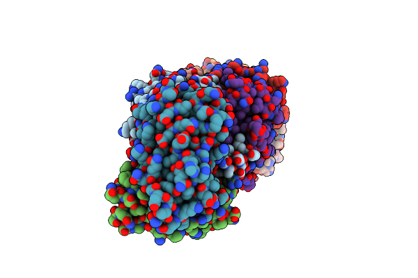
Organism: Shewanella oneidensis mr-1, Escherichia coli
Method: X-RAY DIFFRACTION Resolution:3.08 Å Release Date: 2020-09-30 Classification: ANTITOXIN |
 |
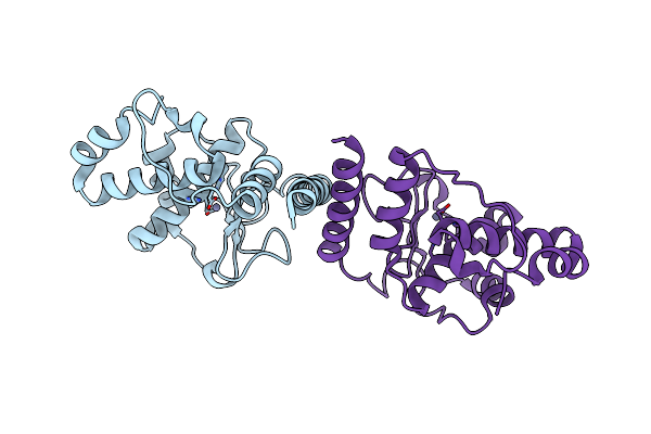
Organism: Shewanella oneidensis mr-1
Method: X-RAY DIFFRACTION Resolution:2.61 Å Release Date: 2020-09-30 Classification: ANTITOXIN |
 |
Organism: Shewanella oneidensis (strain mr-1)
Method: X-RAY DIFFRACTION Resolution:2.77 Å Release Date: 2020-09-30 Classification: TOXIN Ligands: ANP, MG |
 |
High-Resolution Crystal Structure Of Fluoropropylated Cystine Knot, Binding To Alpha-5 Beta-6 Integrin
Organism: Momordica cochinchinensis
Method: X-RAY DIFFRACTION Resolution:1.00 Å Release Date: 2019-08-14 Classification: PROTEIN BINDING |
 |
Organism: Pseudomonas aeruginosa pao1
Method: X-RAY DIFFRACTION Resolution:3.20 Å Release Date: 2019-07-24 Classification: METAL BINDING PROTEIN Ligands: ZN |
 |
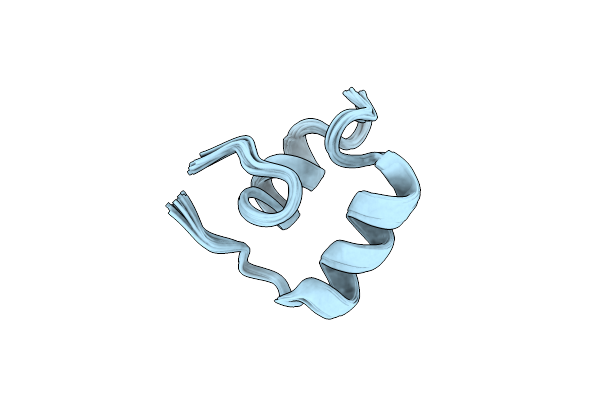
Organism: Ancylostoma caninum
Method: SOLUTION NMR Release Date: 2019-06-19 Classification: IMMUNE SYSTEM |
 |
Structure Of The Chromophore Binding Domain Of Stigmatella Aurantiaca Phytochrome P1, Wild-Type
Organism: Stigmatella aurantiaca dw4/3-1
Method: X-RAY DIFFRACTION Resolution:1.85 Å Release Date: 2018-09-19 Classification: SIGNALING PROTEIN Ligands: BLR |
 |
The Structure Of The Stigmatella Aurantiaca Phytochrome Chromophore Binding Domain T289H Mutant
Organism: Stigmatella aurantiaca dw4/3-1
Method: X-RAY DIFFRACTION Resolution:1.92 Å Release Date: 2018-09-19 Classification: SIGNALING PROTEIN Ligands: BLR |
 |
Organism: Stigmatella aurantiaca dw4/3-1
Method: X-RAY DIFFRACTION Resolution:2.18 Å Release Date: 2018-09-19 Classification: SIGNALING PROTEIN Ligands: BLR |
 |
Organism: Stigmatella aurantiaca dw4/3-1
Method: X-RAY DIFFRACTION Resolution:2.65 Å Release Date: 2018-09-19 Classification: SIGNALING PROTEIN Ligands: BLR |
 |
Stigmatella Aurantiaca Bacterial Phytochrome P1, Pas-Gaf-Phy T289H Mutant, Room Temperature Structure
Organism: Stigmatella aurantiaca dw4/3-1
Method: X-RAY DIFFRACTION Resolution:3.15 Å Release Date: 2018-09-19 Classification: SIGNALING PROTEIN Ligands: BLR |
 |
Organism: Mycobacterium tuberculosis (strain atcc 25618 / h37rv)
Method: X-RAY DIFFRACTION Resolution:2.45 Å Release Date: 2018-06-27 Classification: HYDROLASE Ligands: PO4 |
 |
Beta-Lactamase, Mixed With Ceftriaxone, 30Ms Time Point, Shards Crystal Form
Organism: Mycobacterium tuberculosis (strain atcc 25618 / h37rv)
Method: X-RAY DIFFRACTION Resolution:2.75 Å Release Date: 2018-06-27 Classification: HYDROLASE Ligands: PO4, 9F2 |
 |
Organism: Mycobacterium tuberculosis (strain atcc 25618 / h37rv)
Method: X-RAY DIFFRACTION Resolution:2.15 Å Release Date: 2018-06-27 Classification: Hydrolase/Antibiotic Ligands: PO4, 9F2, FZS |
 |
Organism: Mycobacterium tuberculosis (strain atcc 25618 / h37rv)
Method: X-RAY DIFFRACTION Resolution:2.20 Å Release Date: 2018-06-27 Classification: HYDROLASE/ANTIBIOTIC Ligands: PO4, 9F2, CUG, FZS |