
Search Count: 38
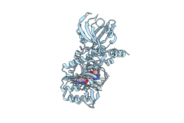 |
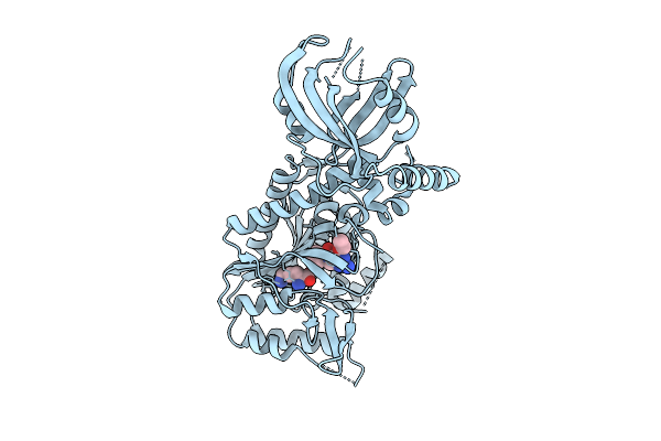
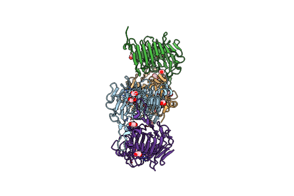
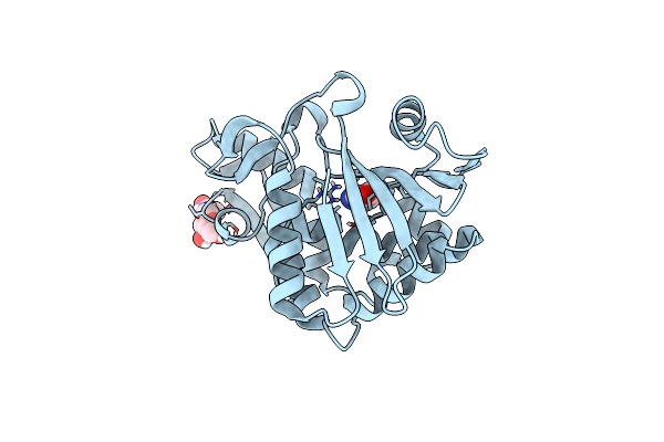
Crystal Structure Of Lysyl-Trna Synthetase From Mycobacterium Tuberculosis Complexed With L-Lysine And Inhibitor Ddd01991231
Organism: Mycobacterium tuberculosis
Method: X-RAY DIFFRACTION Release Date: 2025-08-13 Classification: LIGASE Ligands: LYS, A1I5H |
 |
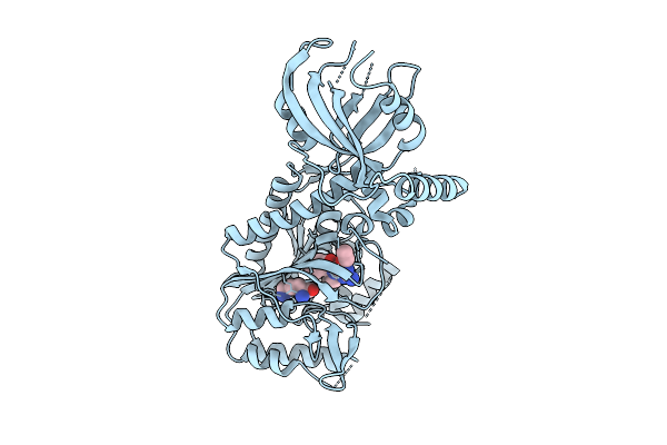
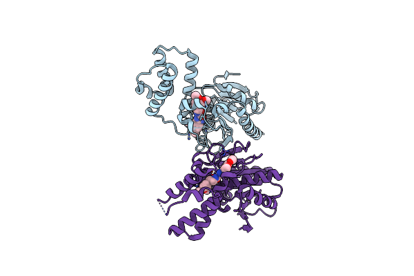
Crystal Structure Of Lysyl-Trna Synthetase From Mycobacterium Tuberculosis Complexed With L-Lysine And Inhibitor Ddd01993593
Organism: Mycobacterium tuberculosis
Method: X-RAY DIFFRACTION Release Date: 2025-08-13 Classification: LIGASE Ligands: LYS, A1I50 |
 |
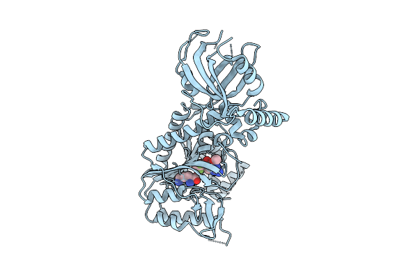
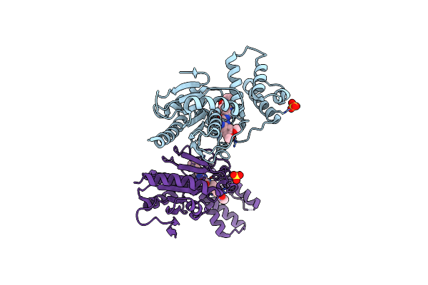
Crystal Structure Of Lysyl-Trna Synthetase From Mycobacterium Tuberculosis Complexed With L-Lysine And Inhibitor Ddd01869767
Organism: Mycobacterium tuberculosis
Method: X-RAY DIFFRACTION Release Date: 2025-08-13 Classification: LIGASE Ligands: LYS, A1I5Z |
 |
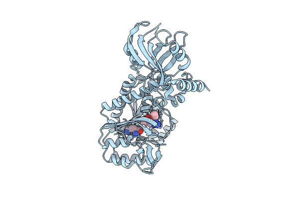
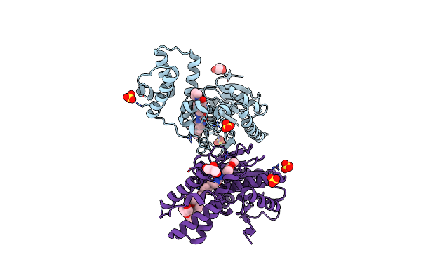
Crystal Structure Of Lysyl-Trna Synthetase From Mycobacterium Tuberculosis Complexed With L-Lysine And Inhibitor Ddd018870911
Organism: Mycobacterium tuberculosis
Method: X-RAY DIFFRACTION Release Date: 2025-08-13 Classification: LIGASE Ligands: LYS, A1I60 |
 |
Crystal Structure Of Lysyl-Trna Synthetase From Mycobacterium Tuberculosis Complexed With L-Lysine And Inhibitor Ddd01839469
Organism: Mycobacterium tuberculosis
Method: X-RAY DIFFRACTION Release Date: 2025-08-13 Classification: LIGASE Ligands: LYS, A1I61 |
 |
Crystal Structure Of Lysyl-Trna Synthetase From Mycobacterium Tuberculosis Complexed With L-Lysine And Inhibitor Ddd01866774
Organism: Mycobacterium tuberculosis
Method: X-RAY DIFFRACTION Release Date: 2025-08-13 Classification: LIGASE Ligands: LYS, A1I62 |
 |
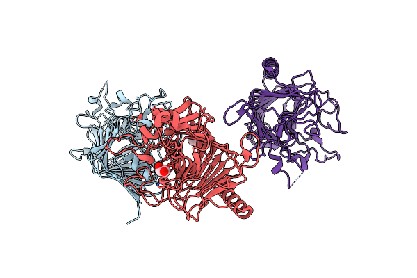
Crystal Structure Of Bacterial Pectin Methylesterase Pmec5 From B. Fibrisolvens D1T
Organism: Butyrivibrio fibrisolvens dsm 3071
Method: X-RAY DIFFRACTION Release Date: 2025-01-15 Classification: SUGAR BINDING PROTEIN Ligands: MG, NA, EDO, NH4 |
 |
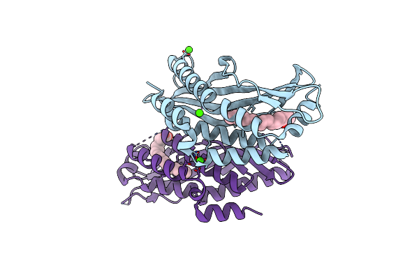
Crystal Structure Of Hydroxyisourate Hydrolase From Herbaspirillum Seropedicae
Organism: Herbaspirillum seropedicae
Method: X-RAY DIFFRACTION Resolution:2.15 Å Release Date: 2024-01-31 Classification: HYDROLASE |
 |
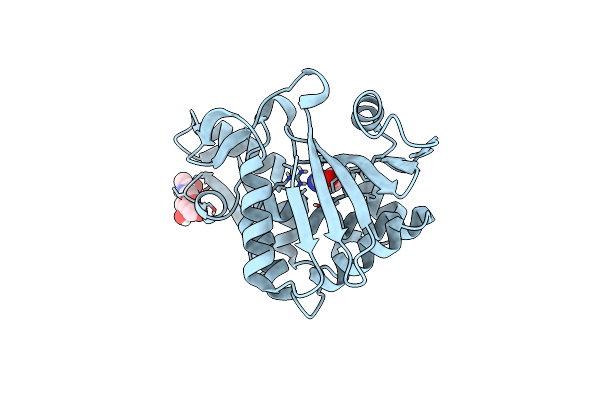
Identification And Optimisation Of Novel Inhibitors Of The Polyketide Synthetase 13 Thioesterase Domain With Antitubercular Activity
Organism: Mycobacterium tuberculosis
Method: X-RAY DIFFRACTION Resolution:1.80 Å Release Date: 2023-11-22 Classification: TRANSFERASE Ligands: IJJ |
 |
Identification And Optimisation Of Novel Inhibitors Of The Polyketide Synthetase 13 Thioesterase Domain With Antitubercular Activity
Organism: Mycobacterium tuberculosis
Method: X-RAY DIFFRACTION Resolution:1.80 Å Release Date: 2023-11-22 Classification: TRANSFERASE Ligands: SO4, IKG |
 |
Identification And Optimisation Of Novel Inhibitors Of The Polyketide Synthetase 13 Thioesterase Domain With Antitubercular Activity
Organism: Mycobacterium tuberculosis
Method: X-RAY DIFFRACTION Resolution:1.71 Å Release Date: 2023-11-22 Classification: TRANSFERASE Ligands: SO4, IL8, GOL, 2Q5 |
 |
Crystal Structure Of Bacterial Pectin Methylesterase Pmec2 From Rumen Butyrivibrio
Organism: Butyrivibrio fibrisolvens
Method: X-RAY DIFFRACTION Resolution:2.30 Å Release Date: 2023-08-16 Classification: SUGAR BINDING PROTEIN Ligands: CL, EDO |
 |
Crystal Structure Of Bacterial Pectin Methylesterase Pme8A From Rumen Butyrivibrio
Organism: Butyrivibrio proteoclasticus
Method: X-RAY DIFFRACTION Resolution:2.30 Å Release Date: 2023-08-16 Classification: SUGAR BINDING PROTEIN Ligands: EDO |
 |
Organism: Bos taurus
Method: X-RAY DIFFRACTION Resolution:2.30 Å Release Date: 2019-03-13 Classification: LIPID BINDING PROTEIN Ligands: CA, OLA |
 |
Organism: Bos taurus
Method: X-RAY DIFFRACTION Resolution:2.00 Å Release Date: 2018-07-11 Classification: LIPID BINDING PROTEIN Ligands: CA |
 |
Cleavage Of Nicotinamide Adenine Dinucleotide By The Ribosome Inactivating Protein Of Momordica Charantia - Enzyme-Nadp+ Co-Crystallisation.
Organism: Momordica charantia
Method: X-RAY DIFFRACTION Resolution:1.52 Å Release Date: 2015-07-22 Classification: HYDROLASE Ligands: NAG, NCA |
 |
Cleavage Of Nicotinamide Adenine Dinucleotides By The Ribosome Inactivating Protein From Momordica Charantia
Organism: Momordica charantia
Method: X-RAY DIFFRACTION Resolution:1.35 Å Release Date: 2015-05-20 Classification: HYDROLASE Ligands: NAG, NCA |
 |
Organism: Butyrivibrio proteoclasticus
Method: X-RAY DIFFRACTION Resolution:1.33 Å Release Date: 2014-10-08 Classification: HYDROLASE Ligands: CA, TRS |
 |
An Acetyl Xylan Esterase (Est2A) From The Rumen Bacterium Butyrivibrio Proteoclasticus.
Organism: Butyrivibrio proteoclasticus b316
Method: X-RAY DIFFRACTION Resolution:2.10 Å Release Date: 2013-02-13 Classification: HYDROLASE Ligands: ACY, GOL |
 |
An Acetyl Xylan Esterase (Est2A) From The Rumen Bacterium Butyrivibrio Proteoclasticus.
Organism: Butyrivibrio proteoclasticus
Method: X-RAY DIFFRACTION Resolution:2.00 Å Release Date: 2013-02-13 Classification: HYDROLASE Ligands: ACY, PEG, CL, GOL |