
Search Count: 11
 |
Organism: Talaromyces marneffei pm1
Method: X-RAY DIFFRACTION Resolution:4.20 Å Release Date: 2019-03-06 Classification: LIPID BINDING PROTEIN Ligands: NI |
 |
Organism: Klebsiella pneumoniae
Method: X-RAY DIFFRACTION Resolution:0.95 Å Release Date: 2018-03-07 Classification: HYDROLASE Ligands: ZN, OH, EDO, SIN |
 |
Organism: Klebsiella pneumoniae
Method: X-RAY DIFFRACTION Resolution:1.55 Å Release Date: 2018-03-07 Classification: HYDROLASE Ligands: BS3, GOL |
 |
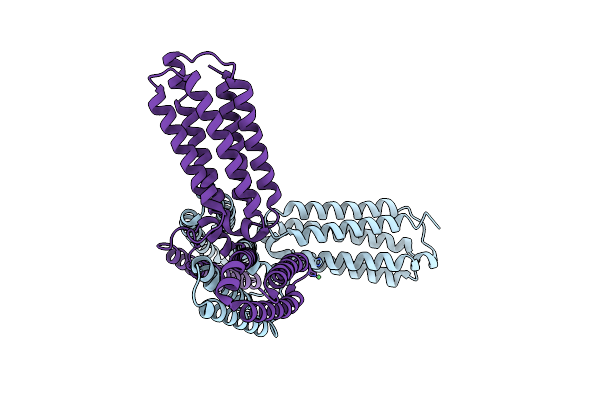
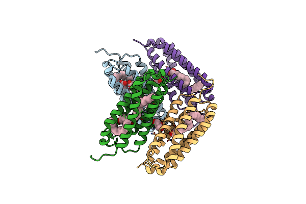
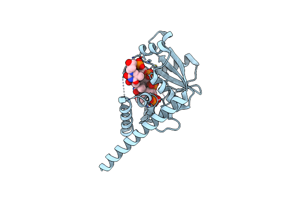
Ligand Binding Domain 2 Of Penicillium Marneffei Mp1 Protein Complexed With Multiple Arachidonic Acids
Organism: Talaromyces marneffei pm1
Method: X-RAY DIFFRACTION Resolution:1.50 Å Release Date: 2016-12-14 Classification: LIPID BINDING PROTEIN Ligands: ACD |
 |
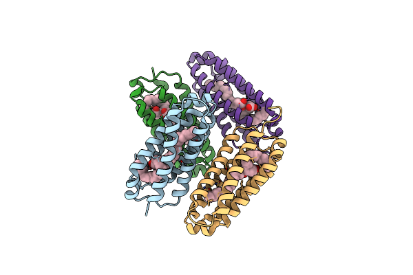
Ligand Binding Domain 1 Of Penicillium Marneffei Mp1 Protein In Complex With Palmitic Acid
Organism: Talaromyces marneffei pm1
Method: X-RAY DIFFRACTION Resolution:1.80 Å Release Date: 2016-10-19 Classification: LIPID BINDING PROTEIN Ligands: PLM, ACT, GOL |
 |
Ligand Binding Domain 1 Of Penicillium Marneffei Mp1 Protein Complexed With Arachidonic Acids
Organism: Talaromyces marneffei pm1
Method: X-RAY DIFFRACTION Resolution:2.60 Å Release Date: 2016-10-19 Classification: LIPID BINDING PROTEIN Ligands: ACD |
 |
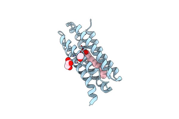
Ligand Binding Domain 2 Of Penicillium Marneffei Mp1 Protein In Complex With Arachidonic Acids
Organism: Talaromyces marneffei pm1
Method: X-RAY DIFFRACTION Resolution:1.45 Å Release Date: 2016-07-27 Classification: LIPID BINDING PROTEIN Ligands: ACD, GOL |
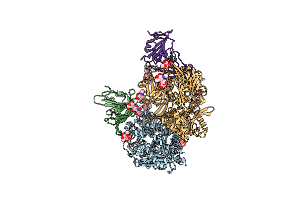 |
Bat-Derived Coronavirus Hku4 Uses Mers-Cov Receptor Human Cd26 For Cell Entry
Organism: Homo sapiens, Tylonycteris bat coronavirus hku4
Method: X-RAY DIFFRACTION Resolution:2.59 Å Release Date: 2014-10-29 Classification: HYDROLASE/VIRAL PROTEIN Ligands: NAG |
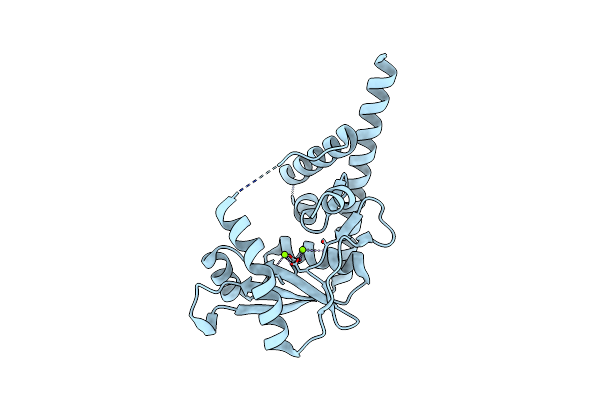 |
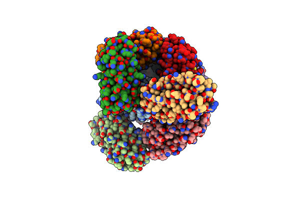
Crystal Structure Of An Alkaline Exonuclease (Lhk-Exo) From Laribacter Hongkongensis
Organism: Laribacter hongkongensis
Method: X-RAY DIFFRACTION Resolution:1.90 Å Release Date: 2012-02-15 Classification: HYDROLASE Ligands: MG |
 |
Organism: Laribacter hongkongensis
Method: X-RAY DIFFRACTION Resolution:2.59 Å Release Date: 2012-02-15 Classification: HYDROLASE Ligands: MG, D5M |
 |
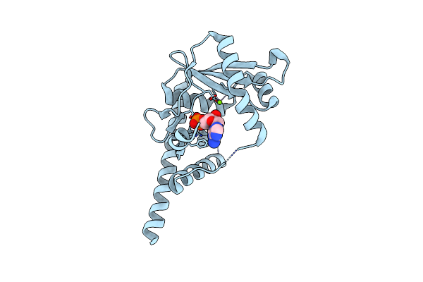
Crystal Structure Of Lhk-Exo In Complex With 5-Phosphorylated Oligothymidine (Dt)4
Organism: Laribacter hongkongensis
Method: X-RAY DIFFRACTION Resolution:2.80 Å Release Date: 2012-02-15 Classification: HYDROLASE/DNA Ligands: MG |