
Search Count: 95
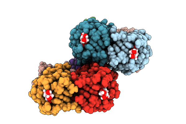 |
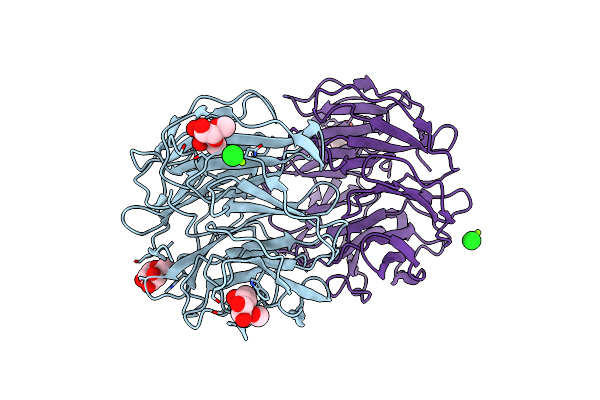
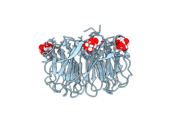
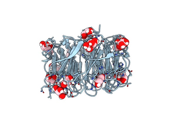
Joint X-Ray/Neutron Room Temperature Structure Of Perdeuterated Leca Lectin In Complex With Deuterated Galactose
Organism: Pseudomonas aeruginosa
Method: X-RAY DIFFRACTION, NEUTRON DIFFRACTION Resolution:1.49 Å, 1.9 Å Release Date: 2025-12-24 Classification: SUGAR BINDING PROTEIN Ligands: GLA, CA |
 |
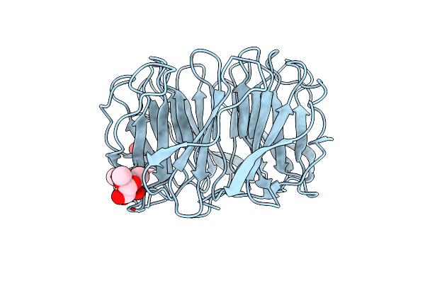
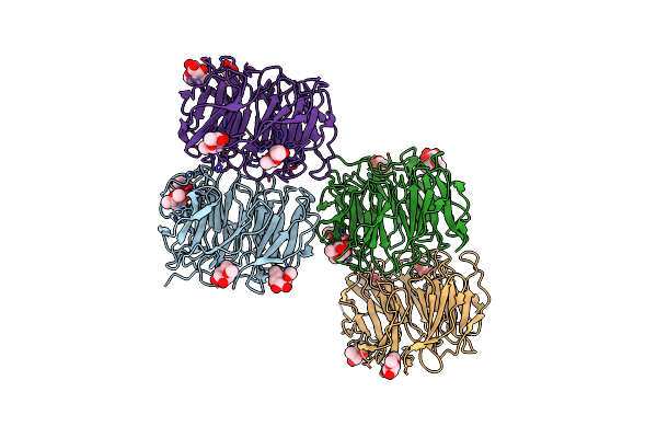
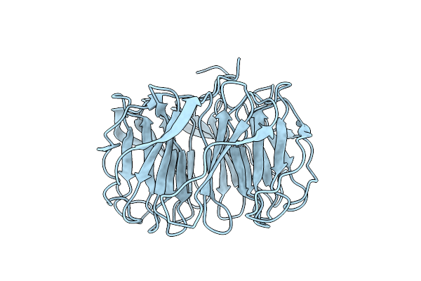
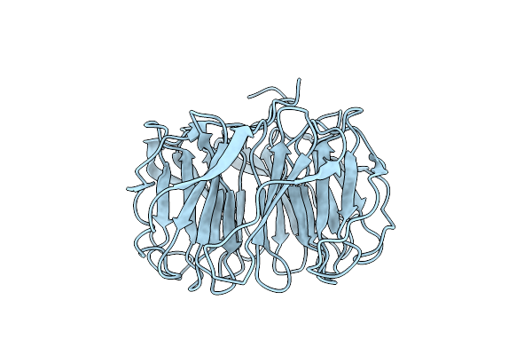
Apo Form Of Tumor Necrosis Factor-Like Lectin Pltl From Photorhabdus Laumondii
Organism: Photorhabdus laumondii subsp. laumondii tto1
Method: X-RAY DIFFRACTION Resolution:1.20 Å Release Date: 2025-10-29 Classification: SUGAR BINDING PROTEIN |
 |
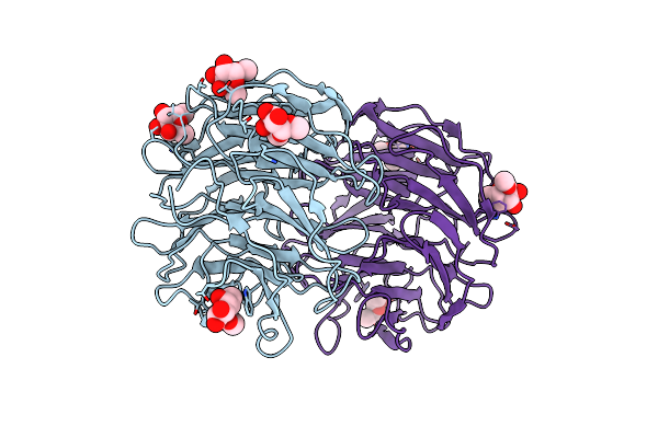
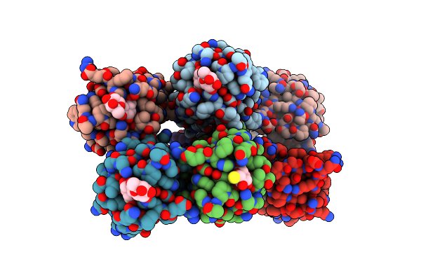
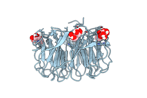
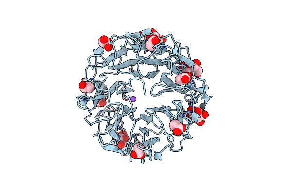
Tumor Necrosis Factor-Like Lectin Pltl From Photorhabdus Laumondii In Complex With Blood Group B Trisaccharide
Organism: Photorhabdus laumondii subsp. laumondii tto1
Method: X-RAY DIFFRACTION Resolution:1.50 Å Release Date: 2025-10-29 Classification: SUGAR BINDING PROTEIN Ligands: FUC, GLA |
 |
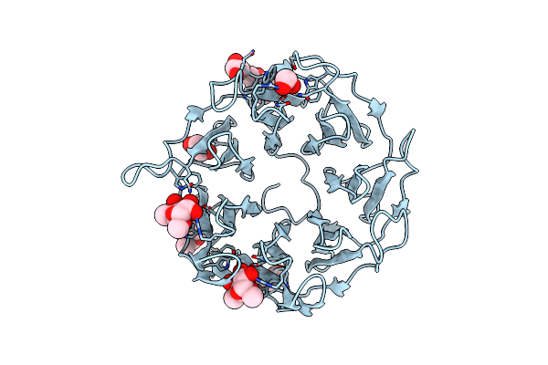
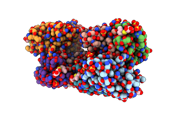
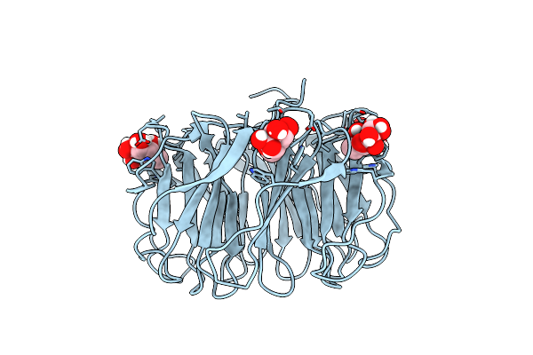
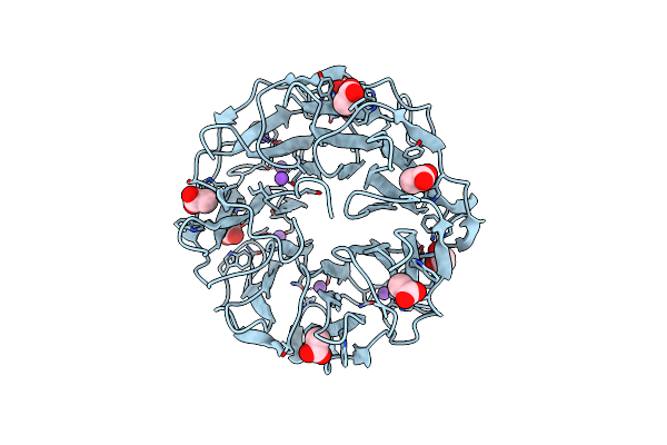
Tumor Necrosis Factor-Like Lectin Pltl From Photorhabdus Laumondii In Complex With Lewis Y Tetrasaccharide
Organism: Photorhabdus laumondii subsp. laumondii tto1
Method: X-RAY DIFFRACTION Resolution:1.60 Å Release Date: 2025-10-29 Classification: SUGAR BINDING PROTEIN |
 |
Tumor Necrosis Factor-Like Lectin Pltl From Photorhabdus Laumondii In Complex With B Lewis B Pentasaccharide
Organism: Photorhabdus laumondii subsp. laumondii tto1
Method: X-RAY DIFFRACTION Resolution:1.30 Å Release Date: 2025-10-29 Classification: SUGAR BINDING PROTEIN |
 |
Organism: Photorhabdus laumondii subsp. laumondii tto1
Method: X-RAY DIFFRACTION Resolution:1.60 Å Release Date: 2025-03-05 Classification: CELL ADHESION Ligands: PEG, MFU |
 |
Organism: Photorhabdus laumondii subsp. laumondii tto1
Method: X-RAY DIFFRACTION Resolution:1.85 Å Release Date: 2025-03-05 Classification: CELL ADHESION Ligands: MFU |
 |
Organism: Photorhabdus laumondii subsp. laumondii tto1
Method: X-RAY DIFFRACTION Resolution:1.50 Å Release Date: 2025-03-05 Classification: CELL ADHESION Ligands: MFU, EDO, PEG |
 |
Organism: Photorhabdus laumondii subsp. laumondii tto1
Method: X-RAY DIFFRACTION Resolution:1.95 Å Release Date: 2025-03-05 Classification: CELL ADHESION Ligands: MFU, CL, MG |
 |
Organism: Photorhabdus laumondii subsp. laumondii tto1
Method: X-RAY DIFFRACTION Resolution:1.70 Å Release Date: 2025-03-05 Classification: CELL ADHESION Ligands: MFU |
 |
Leca From Pseudomonas Aeruginosa In Complex With A Synthetic Thiogalactoside
Organism: Pseudomonas aeruginosa
Method: X-RAY DIFFRACTION Resolution:1.70 Å Release Date: 2024-11-27 Classification: SUGAR BINDING PROTEIN Ligands: CA, CL, YIO, PEG |
 |
Organism: Pseudomonas aeruginosa pao1
Method: X-RAY DIFFRACTION Resolution:1.85 Å Release Date: 2024-11-27 Classification: SUGAR BINDING PROTEIN Ligands: CA, SO4 |
 |
Joint X-Ray/Neutron Room Temperature Structure Of Perdeuterated Pll Lectin In Complex With Perdeuterated L-Fucose
Organism: Photorhabdus laumondii
Method: X-RAY DIFFRACTION, NEUTRON DIFFRACTION Resolution:1.84 Å, 2.2 Å Release Date: 2021-03-24 Classification: SUGAR BINDING PROTEIN Ligands: FUL, FUC |
 |
Room Temperature X-Ray Structure Of Perdeuterated Pll Lectin In Complex With L-Fucose
Organism: Photorhabdus laumondii
Method: X-RAY DIFFRACTION Resolution:1.55 Å Release Date: 2021-03-17 Classification: SUGAR BINDING PROTEIN Ligands: FUL, FUC |
 |
Organism: Photorhabdus laumondii
Method: X-RAY DIFFRACTION Resolution:1.60 Å Release Date: 2021-03-17 Classification: SUGAR BINDING PROTEIN |
 |
Room Temperature X-Ray Structure Of H/D-Exchanged Pll Lectin In Complex With L-Fucose
Organism: Photorhabdus laumondii
Method: X-RAY DIFFRACTION Resolution:1.60 Å Release Date: 2021-03-17 Classification: SUGAR BINDING PROTEIN Ligands: FUC, FUL |
 |
Organism: Photorhabdus laumondii
Method: X-RAY DIFFRACTION Resolution:1.70 Å Release Date: 2021-03-17 Classification: SUGAR BINDING PROTEIN Ligands: FUL, FUC, GOL |
 |
Organism: Photorhabdus laumondii
Method: X-RAY DIFFRACTION, NEUTRON DIFFRACTION Resolution:1.70 Å, 2.20 Å, Release Date: 2021-03-17 Classification: SUGAR BINDING PROTEIN |
 |
Organism: Photorhabdus laumondii subsp. laumondii tto1
Method: X-RAY DIFFRACTION Resolution:1.80 Å Release Date: 2020-07-01 Classification: SUGAR BINDING PROTEIN Ligands: ACT, BGC, EDO, NA |
 |
Organism: Photorhabdus laumondii subsp. laumondii tto1
Method: X-RAY DIFFRACTION Resolution:1.70 Å Release Date: 2020-07-01 Classification: SUGAR BINDING PROTEIN Ligands: ACT, EDO, RM4, NA |