
Search Count: 22
 |
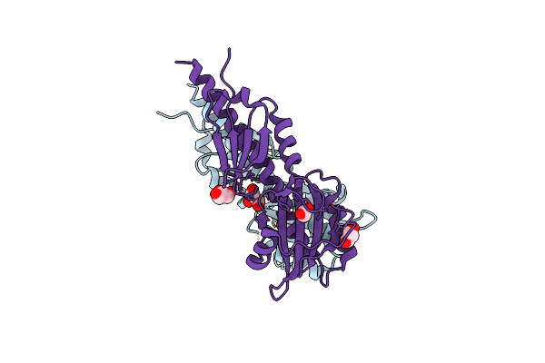
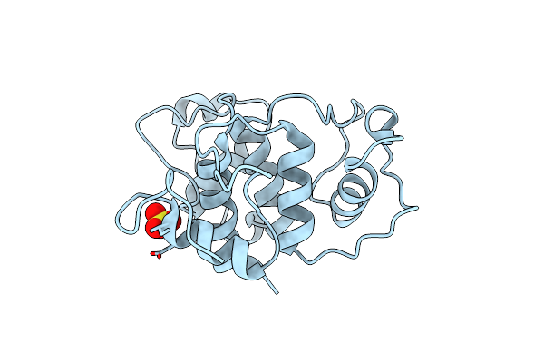
Organism: Mus musculus
Method: X-RAY DIFFRACTION Resolution:2.15 Å Release Date: 2019-11-20 Classification: IMMUNE SYSTEM Ligands: CL, FMT, ACT |
 |
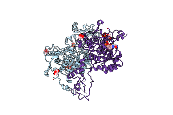
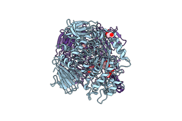
Organism: Mus musculus
Method: X-RAY DIFFRACTION Resolution:2.63 Å Release Date: 2019-11-20 Classification: IMMUNE SYSTEM Ligands: BCG, PEG, GOL, ACT, FMT |
 |
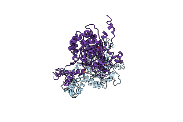
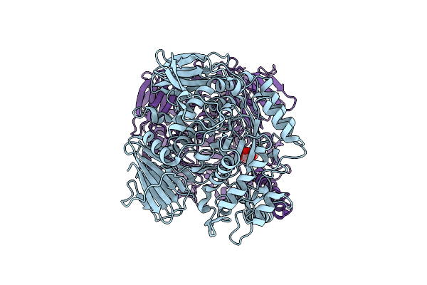
Organism: Anaeromyxobacter dehalogenans
Method: X-RAY DIFFRACTION Resolution:2.00 Å Release Date: 2013-07-17 Classification: SIGNALING PROTEIN Ligands: ZN, ACT |
 |
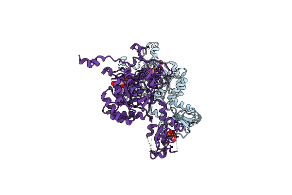
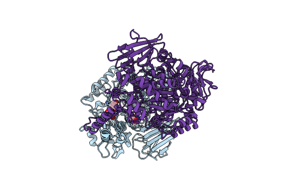
Organism: Anaeromyxobacter dehalogenans
Method: X-RAY DIFFRACTION Resolution:2.00 Å Release Date: 2013-06-12 Classification: SIGNALING PROTEIN Ligands: ZN, CL, NA, ACT |
 |
The Crystal Structure Of Sporulation Kinase D Sensor Domain From Bacillus Subtilis Subsp.
Organism: Bacillus subtilis subsp. subtilis
Method: X-RAY DIFFRACTION Resolution:2.28 Å Release Date: 2013-05-15 Classification: TRANSFERASE Ligands: PYR, GOL |
 |
The Crystal Structure Of Sporulation Kinase D Sensor Domain From Bacillus Subtilis Subsp In Complex With Pyruvate At 2.0A Resolution
Organism: Bacillus subtilis subsp. subtilis
Method: X-RAY DIFFRACTION Resolution:2.03 Å Release Date: 2013-05-15 Classification: TRANSFERASE Ligands: PYR |
 |
The Crystal Structure Of Sporulation Kinase D Mutant Sensor Domain, R131A, From Bacillus Subtilis Subsp In Co-Crystallization With Pyruvate
Organism: Bacillus subtilis subsp. subtilis
Method: X-RAY DIFFRACTION Resolution:2.63 Å Release Date: 2013-05-15 Classification: TRANSFERASE Ligands: ACY |
 |
The Crystal Structure Of Sporulation Kinase D Mutant Sensor Domain, R131A, From Bacillus Subtilis Subsp At 2.4A Resolution
Organism: Bacillus subtilis subsp. subtilis
Method: X-RAY DIFFRACTION Resolution:2.40 Å Release Date: 2013-05-15 Classification: TRANSFERASE Ligands: ACY, GOL |
 |
Organism: Mus musculus
Method: X-RAY DIFFRACTION Resolution:1.80 Å Release Date: 2012-11-14 Classification: IMMUNE SYSTEM Ligands: NHE |
 |
Crystal Structure Of Bacillus Anthracis Inosine Monophosphate Dehydrogenase In The Complex With Imp
Organism: Bacillus anthracis
Method: X-RAY DIFFRACTION Resolution:2.38 Å Release Date: 2011-12-07 Classification: OXIDOREDUCTASE Ligands: IMP, SO4, GOL, CL |
 |
Crystal Structure Of Inosine-5'-Monophosphate Dehydrogenase From Bacillus Anthracis Str. Ames
Organism: Bacillus anthracis
Method: X-RAY DIFFRACTION Resolution:2.60 Å Release Date: 2011-10-05 Classification: OXIDOREDUCTASE Ligands: PO4 |
 |
Crystal Structure Of Inosine-5'-Monophosphate Dehydrogenase From Bacillus Anthracis Str. Ames Complexed With Xmp
Organism: Bacillus anthracis
Method: X-RAY DIFFRACTION Resolution:2.65 Å Release Date: 2011-10-05 Classification: OXIDOREDUCTASE Ligands: XMP, TAR, SO4 |
 |
The Structure Of The Slh Domain From B. Anthracis Surface Array Protein At 1.8A
Organism: Bacillus anthracis
Method: X-RAY DIFFRACTION Resolution:1.80 Å Release Date: 2011-04-27 Classification: STRUCTURAL PROTEIN Ligands: SO4 |
 |
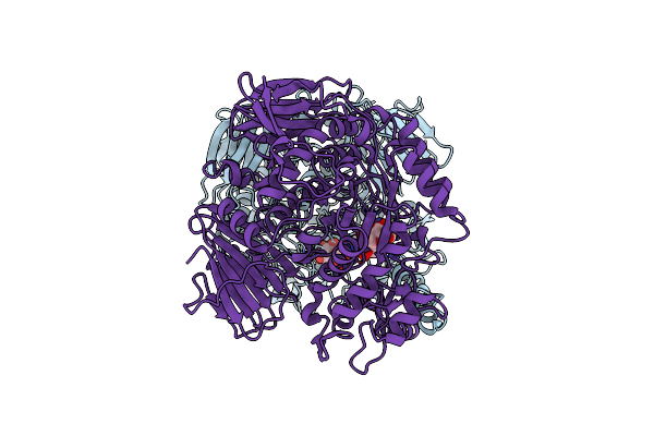
The Crystal Structure Of The D307A Mutant Of Alpha-Glucosidase (Family 31) From Ruminococcus Obeum Atcc 29174 In Complex With Acarbose
Organism: Ruminococcus obeum
Method: X-RAY DIFFRACTION Resolution:1.99 Å Release Date: 2011-01-26 Classification: hydrolase/hydrolase inhibitor Ligands: GLC, AC1, GOL |
 |
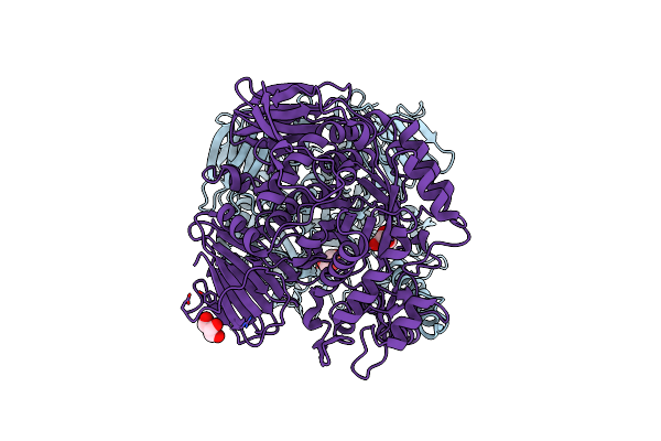
The Crystal Structure Of The W169Y Mutant Of Alpha-Glucosidase (Family 31) From Ruminococcus Obeum Atcc 29174
Organism: Ruminococcus obeum
Method: X-RAY DIFFRACTION Resolution:2.06 Å Release Date: 2010-07-28 Classification: HYDROLASE Ligands: GOL |
 |
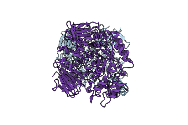
The Crystal Structure Of The The Crystal Structure Of The D420A Mutant Of The Alpha-Glucosidase (Family 31) From Ruminococcus Obeum Atcc 29174
Organism: Ruminococcus obeum
Method: X-RAY DIFFRACTION Resolution:1.57 Å Release Date: 2010-07-21 Classification: HYDROLASE Ligands: TRS |
 |
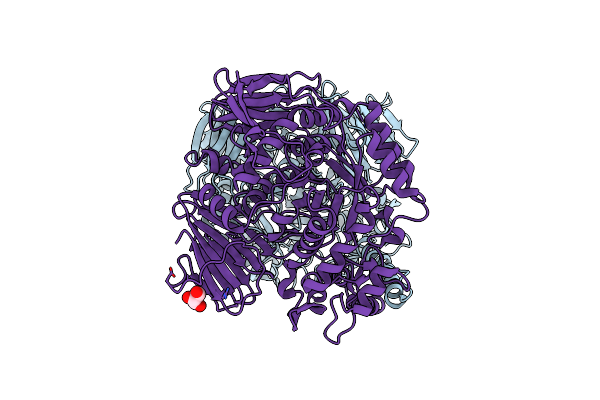
The Crystal Structure Of The D307A Mutant Of Glycoside Hydrolase (Family 31) From Ruminococcus Obeum Atcc 29174 In Complex With Isomaltose
Organism: Ruminococcus obeum
Method: X-RAY DIFFRACTION Resolution:1.91 Å Release Date: 2010-06-23 Classification: HYDROLASE Ligands: GLC, BGC |
 |
The Crystal Structure Of The Alpha-Glucosidase (Family 31) From Ruminococcus Obeum Atcc 29174
Organism: Ruminococcus obeum
Method: X-RAY DIFFRACTION Resolution:2.02 Å Release Date: 2010-06-23 Classification: HYDROLASE Ligands: GOL |
 |
The Crystal Structure Of The D307A Mutant Of Glycoside Hydrolase (Family 31) From Ruminococcus Obeum Atcc 29174
Organism: Ruminococcus obeum
Method: X-RAY DIFFRACTION Resolution:2.90 Å Release Date: 2010-04-21 Classification: HYDROLASE |
 |
The Crystal Structure Of The D73A Mutant Of Glycoside Hydrolase (Family 31) From Ruminococcus Obeum Atcc 29174
Organism: Ruminococcus obeum
Method: X-RAY DIFFRACTION Resolution:2.66 Å Release Date: 2010-03-23 Classification: HYDROLASE Ligands: GOL |