
Search Count: 62
 |
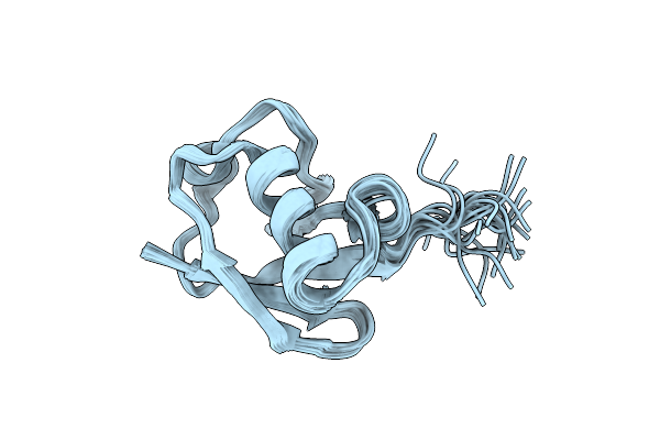
Organism: Mus musculus
Method: SOLUTION NMR Release Date: 2023-05-24 Classification: SIGNALING PROTEIN |
 |
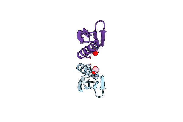
Organism: Homo sapiens
Method: SOLUTION NMR Release Date: 2019-10-30 Classification: STRUCTURAL PROTEIN |
 |
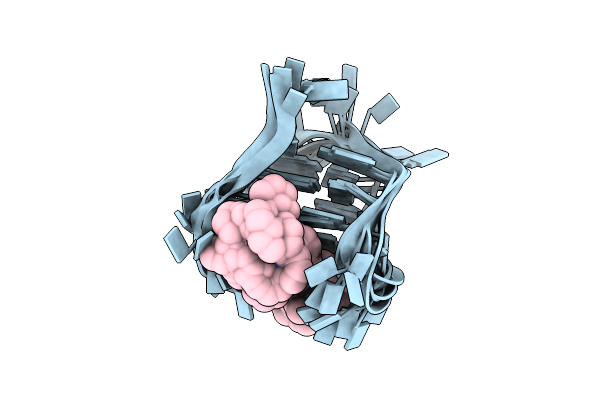
Organism: Staphylococcus simulans
Method: X-RAY DIFFRACTION Resolution:2.50 Å Release Date: 2019-10-16 Classification: PEPTIDE BINDING PROTEIN Ligands: K5T |
 |
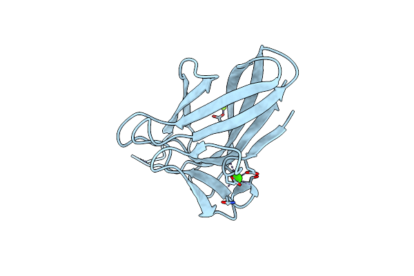
Organism: Staphylococcus simulans
Method: X-RAY DIFFRACTION Resolution:1.43 Å Release Date: 2019-10-16 Classification: PEPTIDE BINDING PROTEIN Ligands: K5T, EDO |
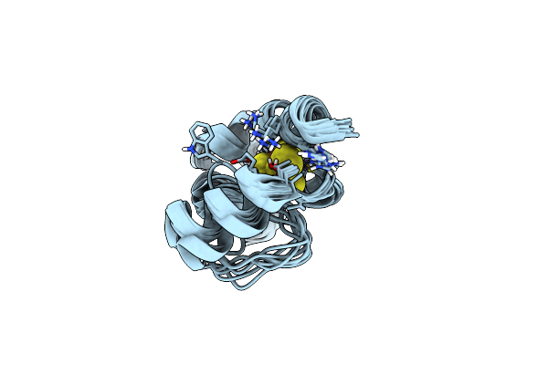 |
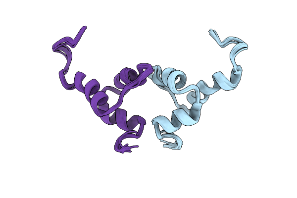
M. Tuberculosis [4Fe-4S] Protein Whib1 Is A Four-Helix Bundle That Forms A No-Sensitive Complex With Sigmaa And Regulates The Major Virulence Factor Esx-1
Organism: Mycobacterium tuberculosis (strain atcc 25618 / h37rv)
Method: SOLUTION NMR Release Date: 2018-01-03 Classification: SIGNALING PROTEIN Ligands: SF4 |
 |
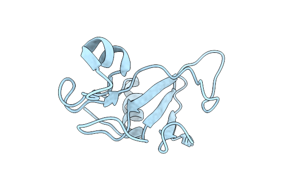
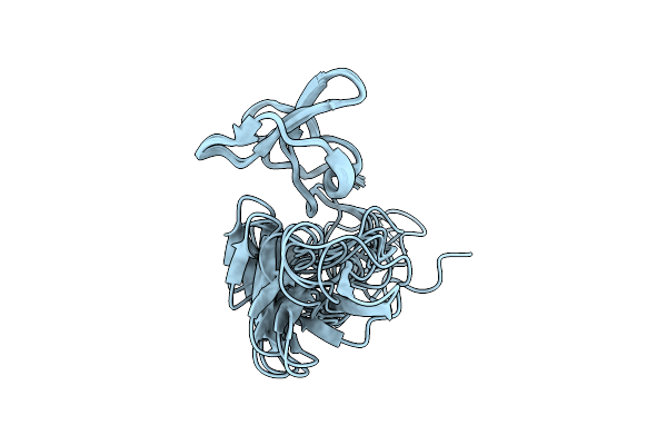
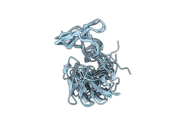
Solution Structure Of Lysm The Peptidoglycan Binding Domain Of Autolysin Atla From Enterococcus Faecalis
Organism: Enterococcus faecalis
Method: SOLUTION NMR Release Date: 2014-06-18 Classification: HYDROLASE |
 |
Organism: Homo sapiens
Method: SOLUTION NMR Release Date: 2014-02-12 Classification: PROTEIN BINDING |
 |
Structural Studies On Dinuclear Ruthenium(Ii) Complexes That Bind Diastereoselectively To An Anti-Parallel Folded Human Telomere Sequence
|
 |
Structural Studies On Dinuclear Ruthenium(Ii) Complexes That Bind Diastereoselectively To An Anti-Parallel Folded Human Telomere Sequence
|
 |
Organism: Homo sapiens
Method: SOLUTION NMR Release Date: 2013-03-27 Classification: PROTEIN BINDING |
 |
Organism: Streptococcus sp. 'group g'
Method: X-RAY DIFFRACTION Resolution:1.20 Å Release Date: 2011-02-23 Classification: PROTEIN BINDING Ligands: FMT |
 |
Organism: Clostridium thermocellum
Method: X-RAY DIFFRACTION Resolution:1.50 Å Release Date: 2010-07-14 Classification: SUGAR BINDING PROTEIN Ligands: CA, MG |
 |
Organism: Homo sapiens
Method: SOLUTION NMR Release Date: 2009-12-15 Classification: PROTEIN BINDING |
 |
Organism: Bacillus amyloliquefaciens
Method: SOLUTION NMR Release Date: 2009-12-08 Classification: HYDROLASE |
 |
Organism: Bacillus amyloliquefaciens
Method: SOLUTION NMR Release Date: 2009-12-08 Classification: HYDROLASE |
 |
Organism: Bacillus amyloliquefaciens
Method: SOLUTION NMR Release Date: 2009-12-08 Classification: HYDROLASE |
 |
Organism: Bacillus amyloliquefaciens
Method: SOLUTION NMR Release Date: 2009-12-08 Classification: HYDROLASE |
 |
Organism: Piromyces equi
Method: SOLUTION NMR Release Date: 2007-09-25 Classification: PROTEIN BINDING |
 |
Organism: Piromyces equi
Method: SOLUTION NMR Release Date: 2007-09-25 Classification: PROTEIN BINDING |
 |
Organism: Streptococcus sp.
Method: SOLUTION NMR Release Date: 2007-09-25 Classification: IMMUNOGLOBULIN |