
Search Count: 20
 |
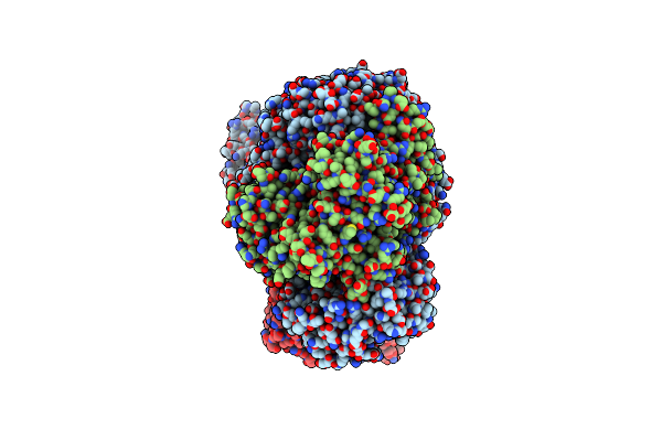
Organism: Photorhabdus thracensis, Escherichia coli
Method: ELECTRON MICROSCOPY Release Date: 2025-03-26 Classification: DNA BINDING PROTEIN Ligands: MG, ATP |
 |
Organism: Photorhabdus thracensis, Escherichia coli
Method: ELECTRON MICROSCOPY Release Date: 2025-03-26 Classification: DNA BINDING PROTEIN Ligands: MG, ATP |
 |
Organism: Photorhabdus thracensis, Escherichia coli
Method: ELECTRON MICROSCOPY Release Date: 2025-03-26 Classification: DNA BINDING PROTEIN Ligands: MG, ATP |
 |
Organism: Photorhabdus thracensis, Escherichia coli
Method: ELECTRON MICROSCOPY Release Date: 2025-03-26 Classification: DNA BINDING PROTEIN Ligands: MG, ATP |
 |
Organism: Photorhabdus thracensis, Escherichia coli
Method: ELECTRON MICROSCOPY Release Date: 2025-03-26 Classification: DNA BINDING PROTEIN Ligands: MG, ATP |
 |
Organism: Escherichia coli, Escherichia phage t7
Method: ELECTRON MICROSCOPY Release Date: 2025-03-26 Classification: DNA BINDING PROTEIN |
 |
Organism: Escherichia coli, Escherichia phage t7
Method: ELECTRON MICROSCOPY Release Date: 2025-03-26 Classification: DNA BINDING PROTEIN |
 |
Organism: Escherichia coli, Escherichia phage t7
Method: ELECTRON MICROSCOPY Release Date: 2022-12-28 Classification: DNA BINDING PROTEIN Ligands: MG |
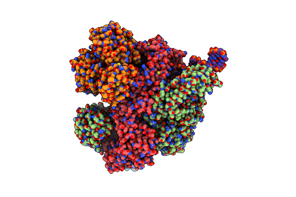 |
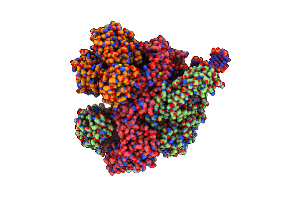
Organism: Escherichia coli, Salmonella phage p22, Synthetic construct
Method: ELECTRON MICROSCOPY Release Date: 2022-12-28 Classification: DNA BINDING PROTEIN Ligands: ANP, MG |
 |
Organism: Escherichia coli, Salmonella phage p22, Synthetic construct
Method: ELECTRON MICROSCOPY Release Date: 2022-12-28 Classification: DNA BINDING PROTEIN Ligands: ANP, MG |
 |
Crystal Structure Of The N-Terminal Domain Of Burkholderia Pseudomallei Antitoxin Hicb
Organism: Burkholderia pseudomallei k96243
Method: X-RAY DIFFRACTION Resolution:1.56 Å Release Date: 2018-10-31 Classification: ANTITOXIN |
 |
Organism: Burkholderia pseudomallei
Method: X-RAY DIFFRACTION Resolution:1.85 Å Release Date: 2018-10-31 Classification: ANTITOXIN Ligands: CL, GOL |
 |
Organism: Burkholderia pseudomallei k96243
Method: X-RAY DIFFRACTION Resolution:2.49 Å Release Date: 2018-10-31 Classification: ANTITOXIN Ligands: SO4, EDO, PGE |
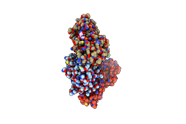 |
Crystal Structure Of The Brct Domains Of 53Bp1 In Complex With P53 And H2Ax-Pser139 (Gammah2Ax)
Organism: Homo sapiens
Method: X-RAY DIFFRACTION Resolution:3.00 Å Release Date: 2015-12-16 Classification: ANTITUMOR PROTEIN Ligands: ZN |
 |
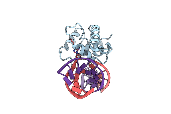
Crystal Structure Of S. Pombe Atl1 In Complex With Damaged Dna Containing 2-Aminopurine
Organism: Schizosaccharomyces pombe
Method: X-RAY DIFFRACTION Resolution:2.85 Å Release Date: 2012-12-26 Classification: DNA BINDING PROTEIN/DNA |
 |
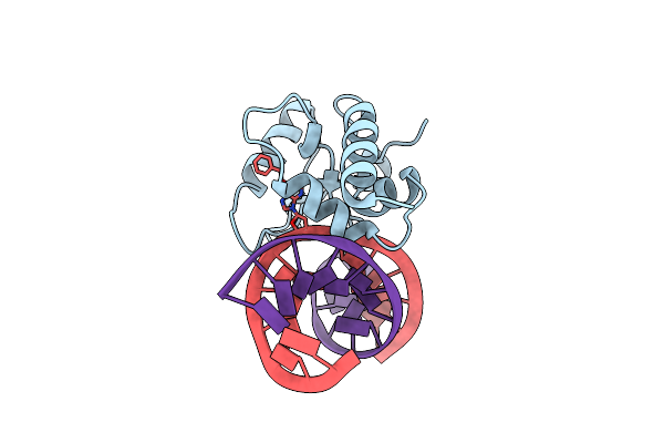
Crystal Structure Of S. Pombe Atl1 In Complex With Damaged Dna Containing 2,6-Diaminopurine
Organism: Schizosaccharomyces pombe
Method: X-RAY DIFFRACTION Resolution:2.70 Å Release Date: 2012-12-26 Classification: DNA BINDING PROTEIN/DNA Ligands: HOH |
 |
Crystal Structure Of S. Pombe Atl1 In Complex With Damaged Dna Containing O6-Hydroxyethylguanine
Organism: Schizosaccharomyces pombe, Synthetic construct
Method: X-RAY DIFFRACTION Resolution:3.10 Å Release Date: 2012-06-20 Classification: DNA BINDING PROTEIN/DNA |
 |
Crystal Structure Of S. Pombe Atl1 In Complex With Damaged Dna Containing O6-Propylguanine
Organism: Schizosaccharomyces pombe, Synthetic construct
Method: X-RAY DIFFRACTION Resolution:3.04 Å Release Date: 2012-06-20 Classification: DNA BINDING PROTEIN/DNA Ligands: HOH |
 |
Crystal Structure Of S. Pombe Atl1 In Complex With Damaged Dna Containing O6-Benzylguanine
Organism: Schizosaccharomyces pombe, Synthetic construct
Method: X-RAY DIFFRACTION Resolution:2.84 Å Release Date: 2012-06-20 Classification: DNA BINDING PROTEIN/DNA |
 |
Crystal Structure Of S. Pombe Atl1 In Complex With Damaged Dna Containing O6-Carboxymethylguanine
Organism: Schizosaccharomyces pombe 972h-, Synthetic construct
Method: X-RAY DIFFRACTION Resolution:2.84 Å Release Date: 2012-06-20 Classification: DNA BINDING PROTEIN/DNA |